The winners of this year’s Nikon Small World Competition have been announced, showcasing the best microphotography that 2021 had to offer.
This is the 46th running of the annual competition, which recognises the year’s most outstanding microphotography across a wide range of scientific disciplines. A panel of judges selected 20 winning entries, and also images worthy of honourable mention and distinction.
Trichome and stomata
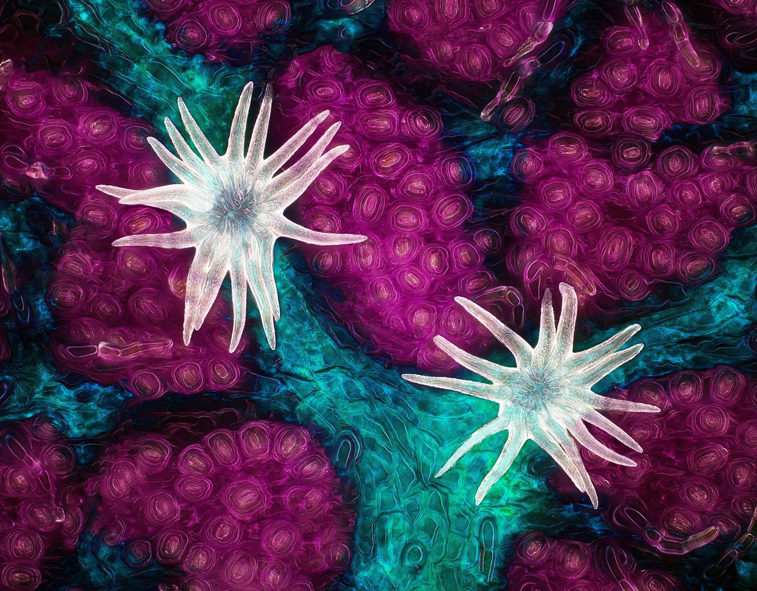
Biologist Jason Kirk from Baylor College of Medicine was awarded first place for this image of hair-like trichomes (shown in white) and stomata (the purple pores), as seen on a southern live oak leaf.
Your brain on microfluidics
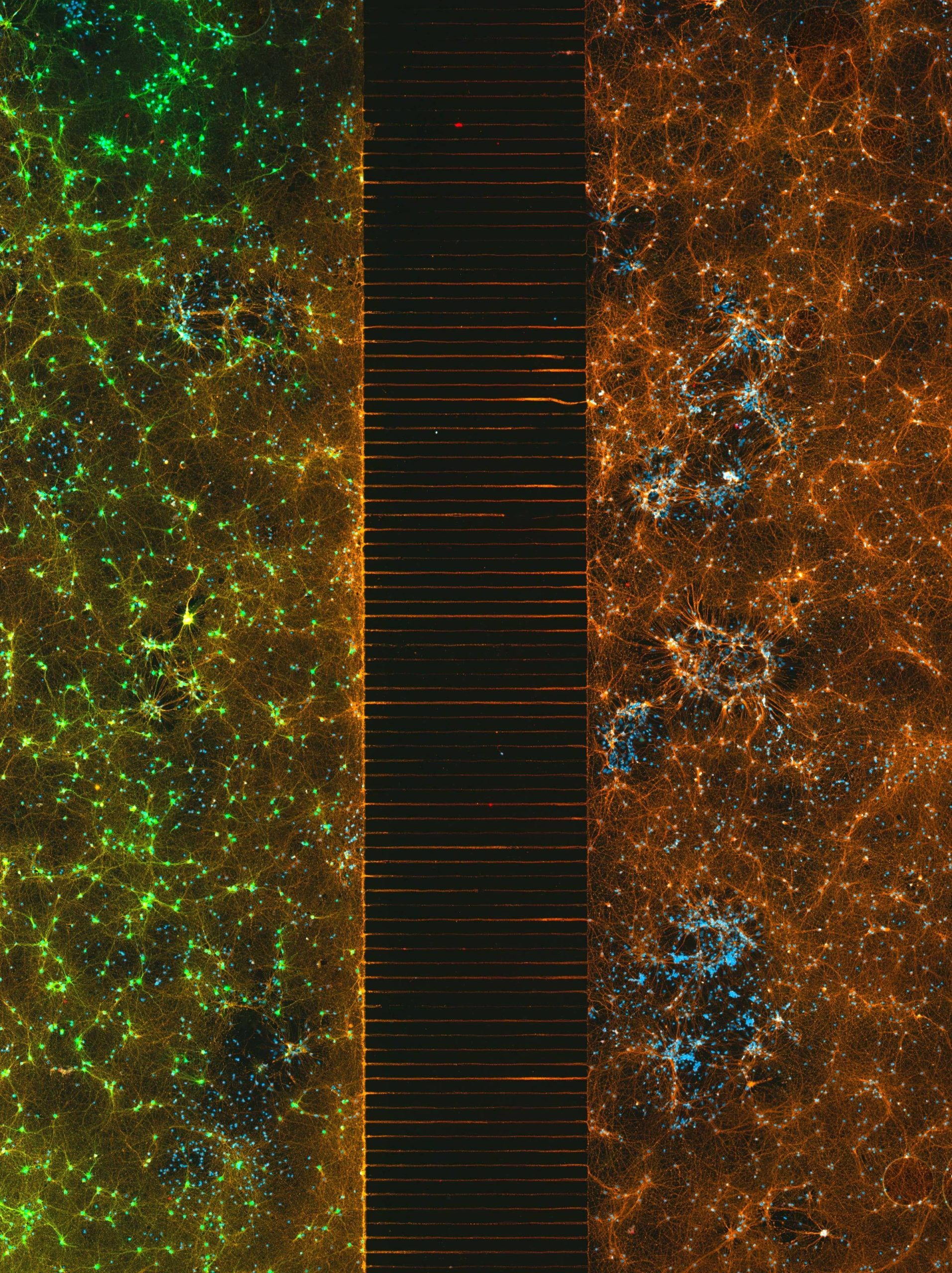
Second place went to Esmeralda Paric and Holly Stefen from the Dementia Research Centre at Macquarie Park in Australia. Their image, colorized with fluorescence, shows a microfluidic device (a tool for managing the flow of liquids inside microscopic-sized channels) consisting of 300,000 networking neurons in two isolated brain samples.
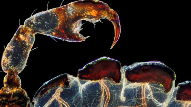
Zoomed-in louse
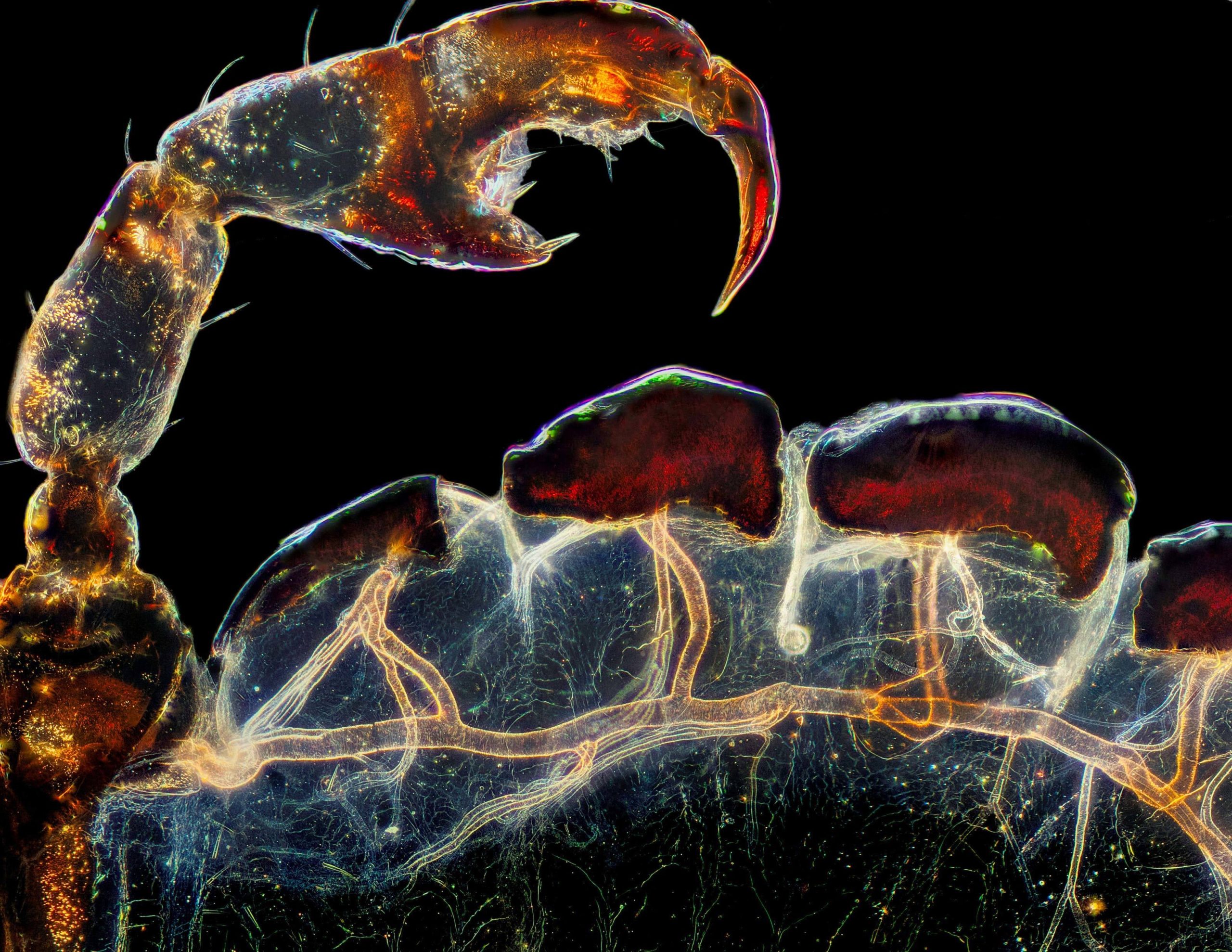
This stunning image earned Frank Reiser of Nassau Community College the third-place prize in this year’s competition. The image, at five-times magnification, shows the rear leg, claw, and respiratory trachea of a louse (the singular for lice).
A sensory neuron
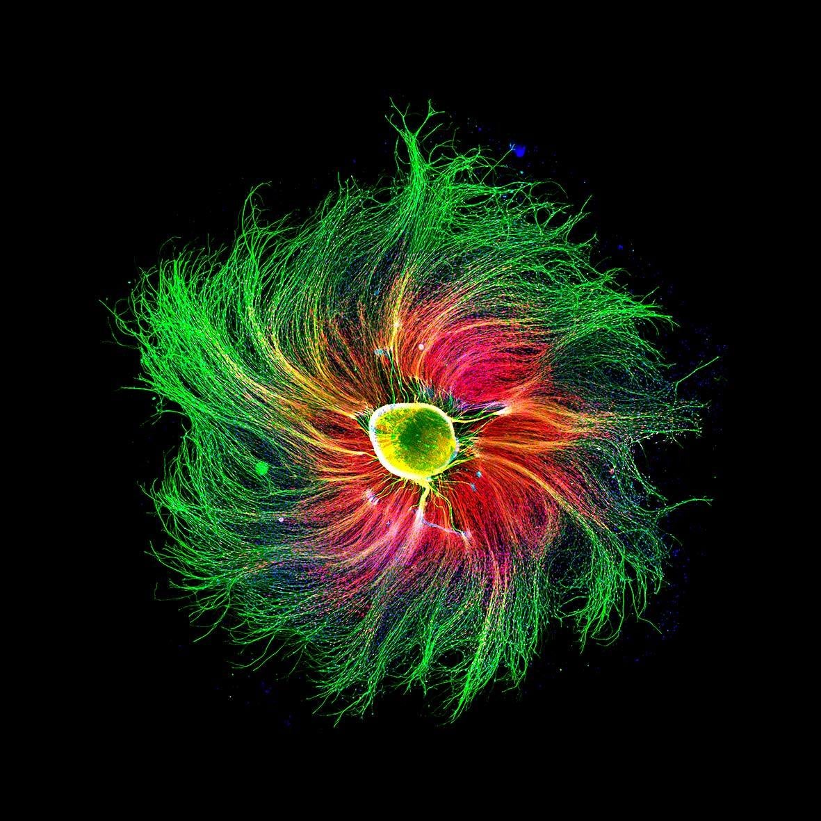
Using fluorescence, Paula Diaz from Pontificia Universidad Católica de Chile was able to show the finer details of a sensory neuron from an embryonic rat. The fourth-place image is shown as 10-times magnification.
Mouth of a fly
This alien-like appendage is the proboscis, or mouth, of a common housefly (Musca domestica) shown at 40-times magnification. The fifth-place image was captured by Oliver Dum from Medienbunker Produktion in Germany.
Blood vessels in a mouse brain
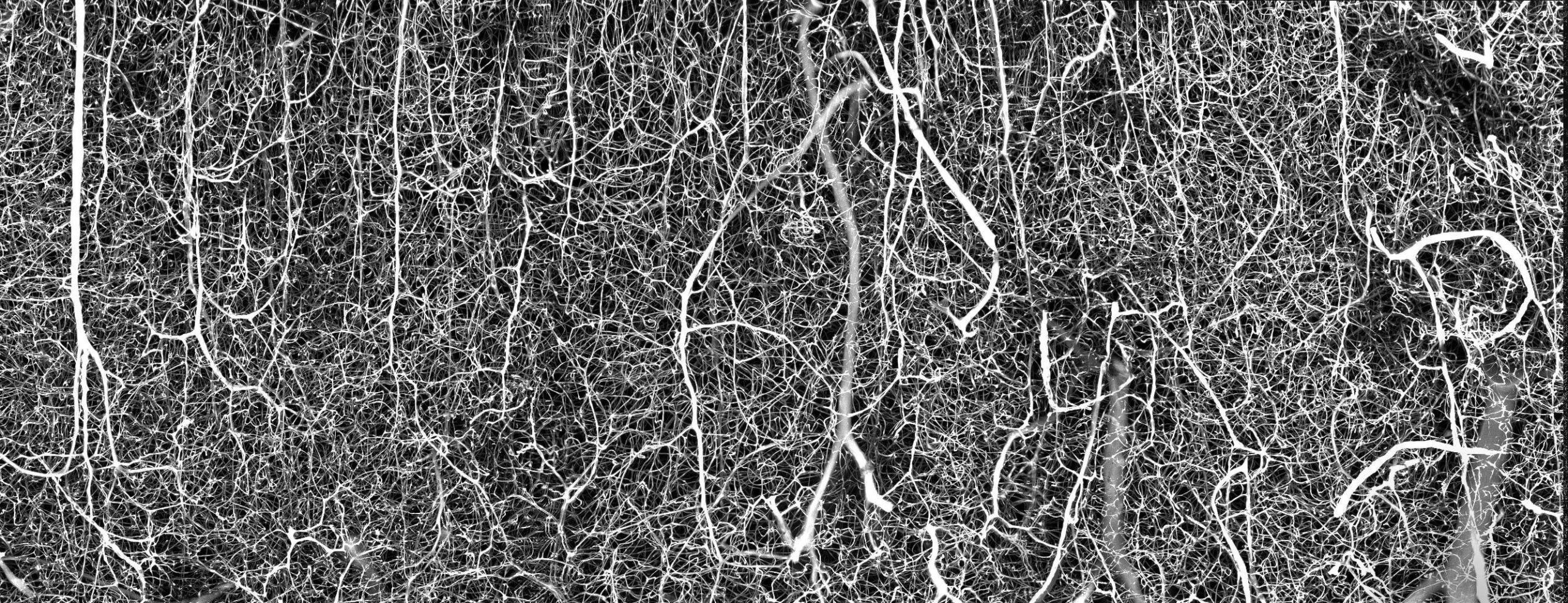
Andrea Tedeschi of The Ohio State University won the sixth-place prize for an image showing the intricate — if not chaotic — three-dimensional vasculature of an adult mouse brain.
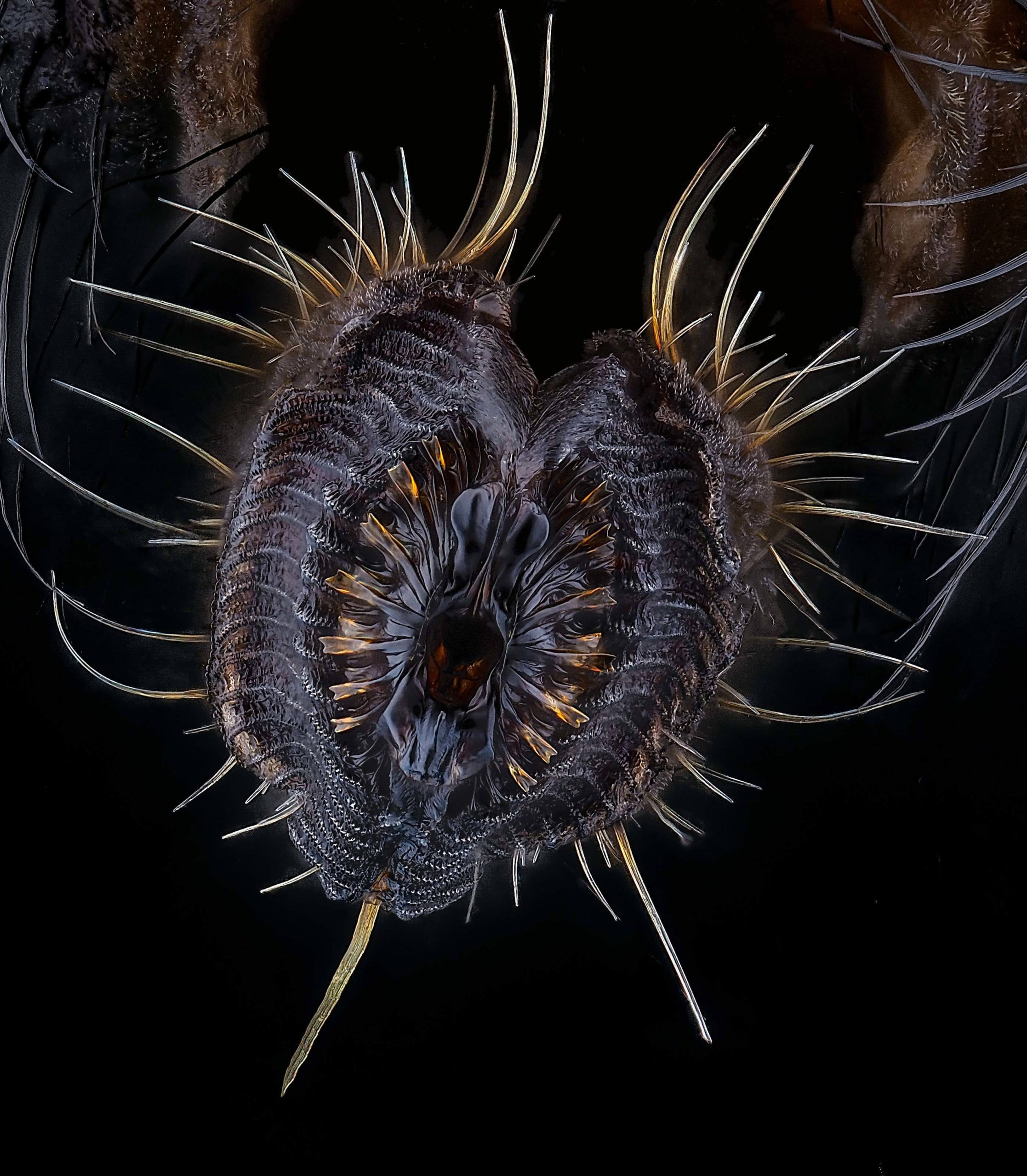
Tick head
This seventh-place image, shown at 10-times magnification, reveals the weirdness that is a tick head.
Mouse guts
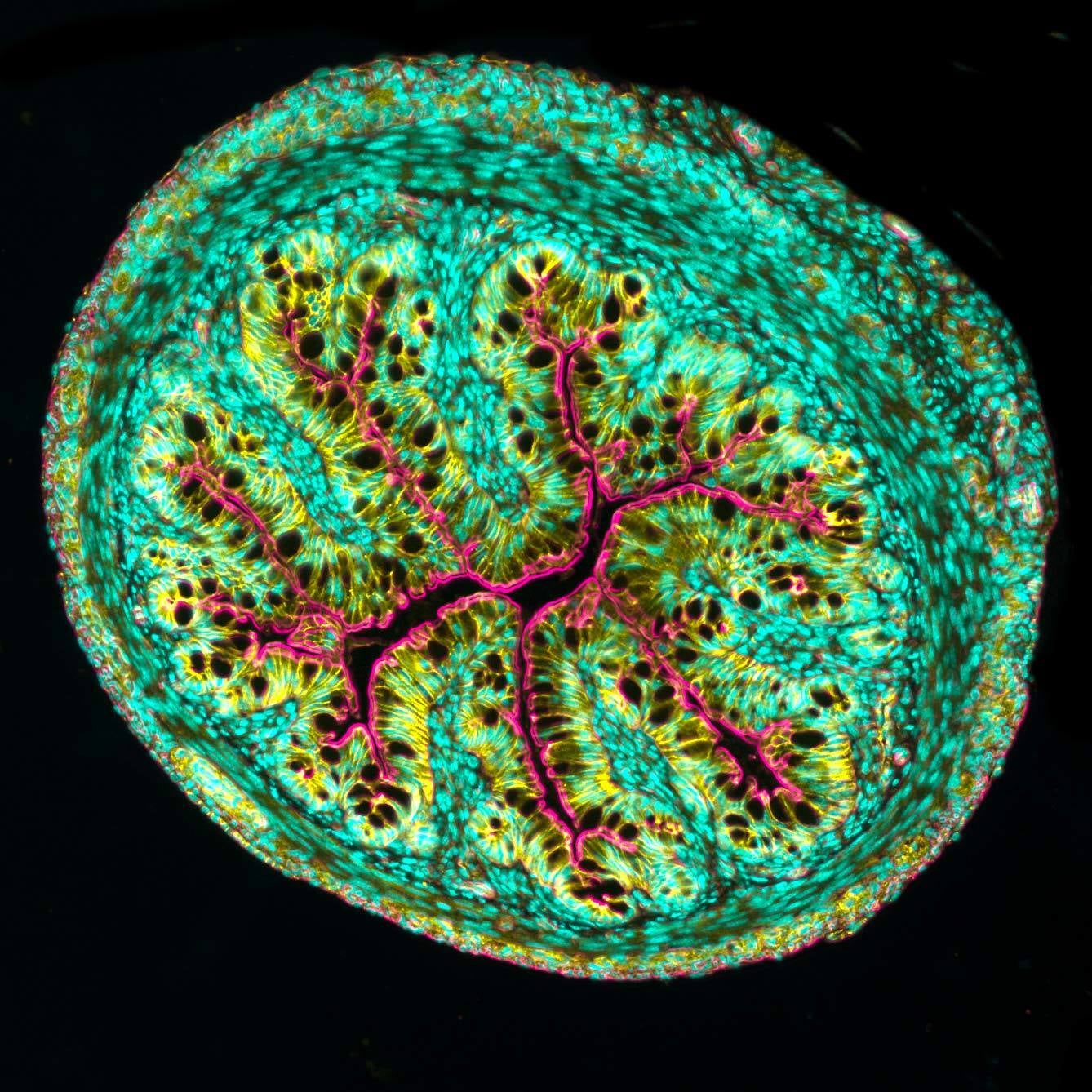
A cross-section section of a mouse intestine, as imaged by Amy Engevik from the Department of Regenerative Medicine and Cell Biology at Medical University of South Carolina.
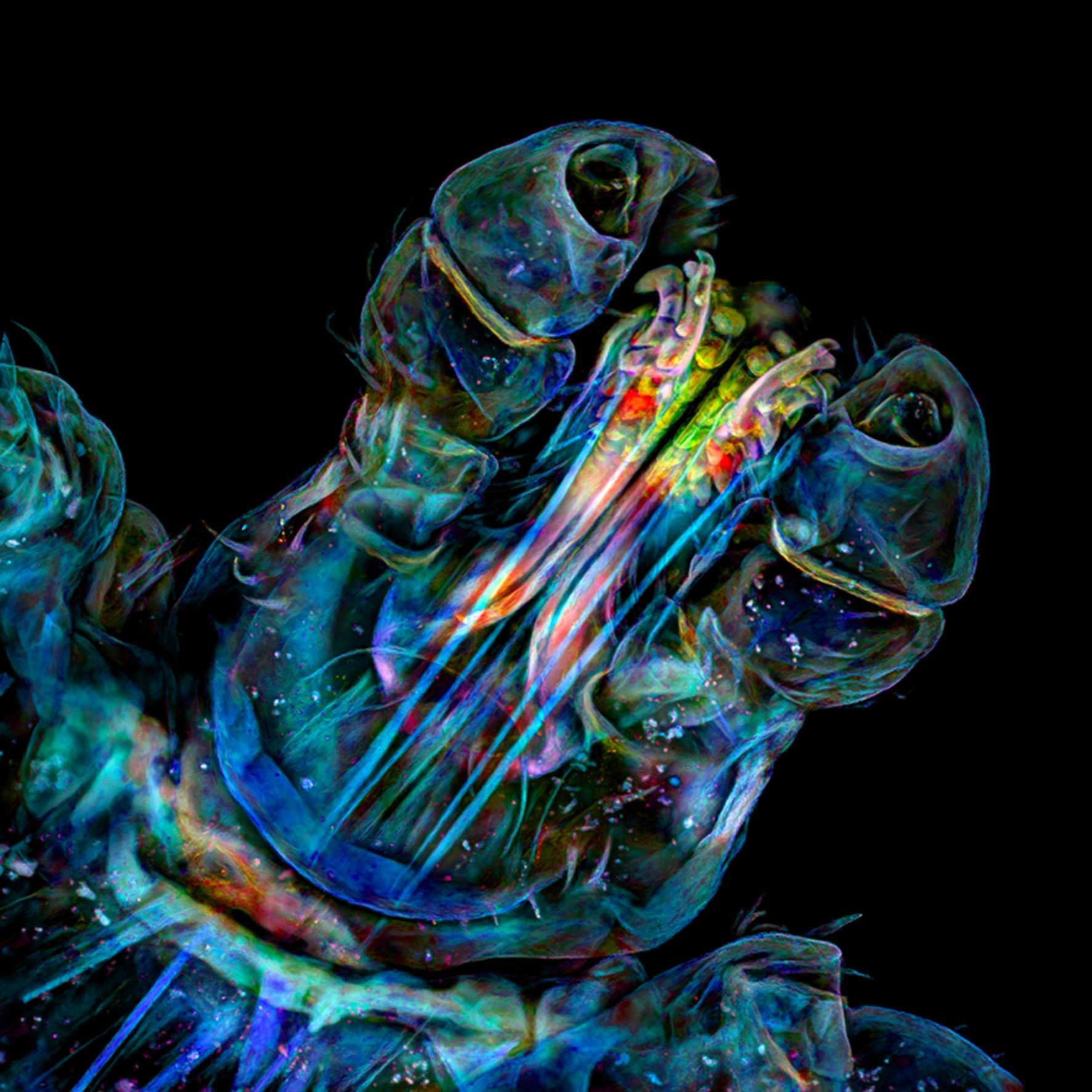
Pregnant water flea
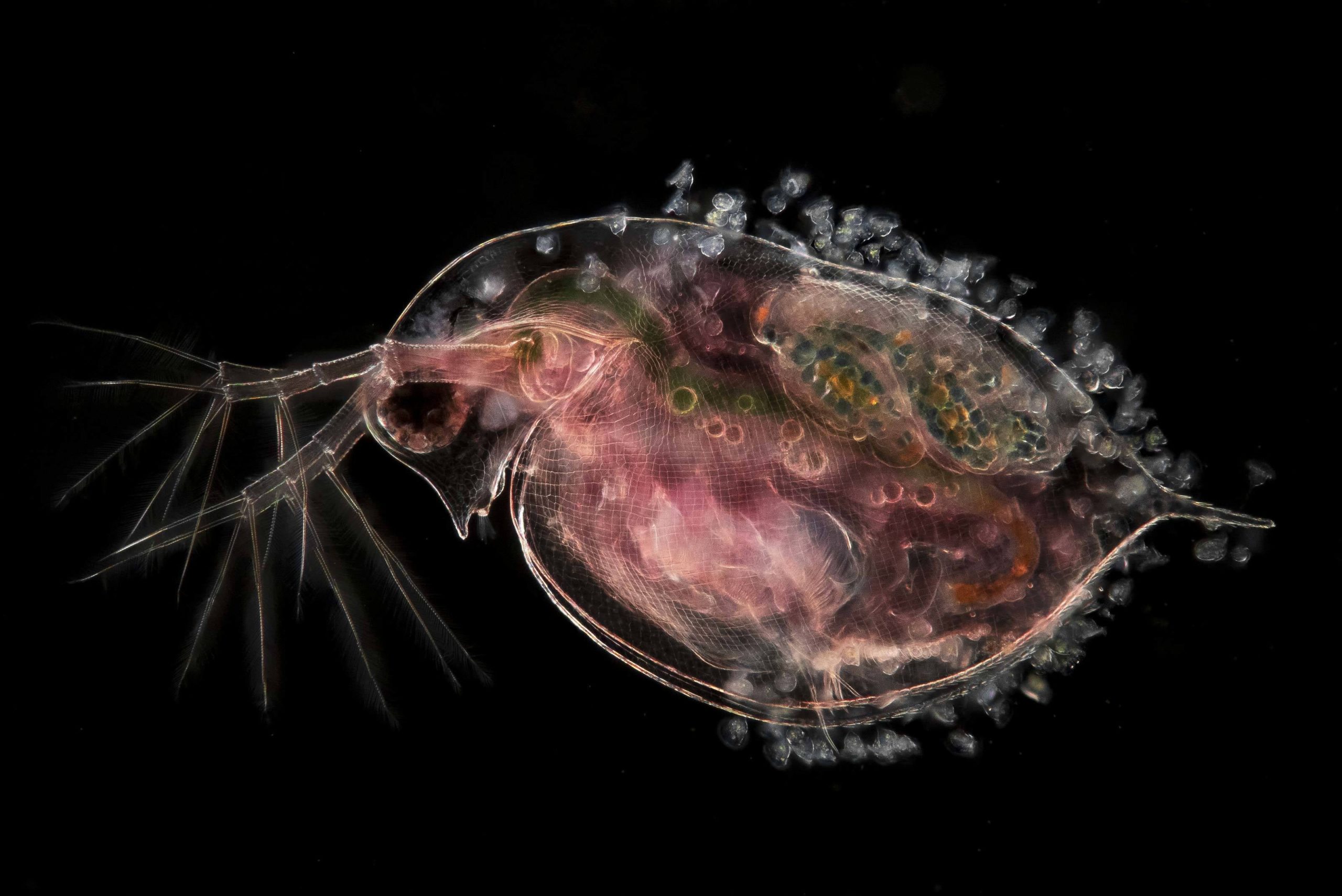
A pregnant water flea carrying embryos and parasitic peritrichs (the white-coloured growths shown along its exterior portions). Jan van IJken of The Netherlands was awarded ninth place for this very cool image.
Butterfly wing
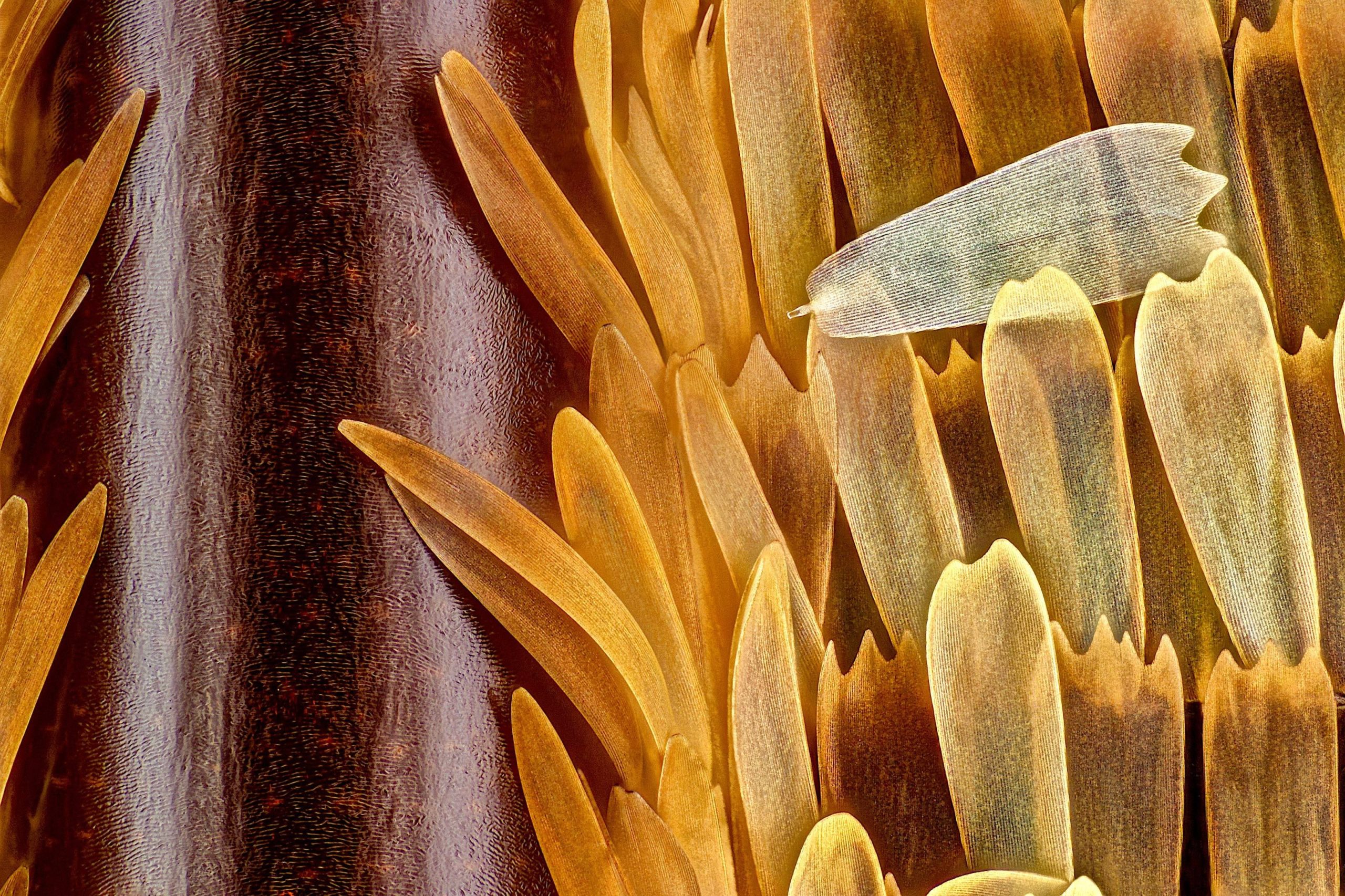
Sébastien Malo of France won the 10th-place prize for this close-up view of a butterfly wing (Morpho didius), showing veins and scales.
Blood vessels in the eye of a mouse
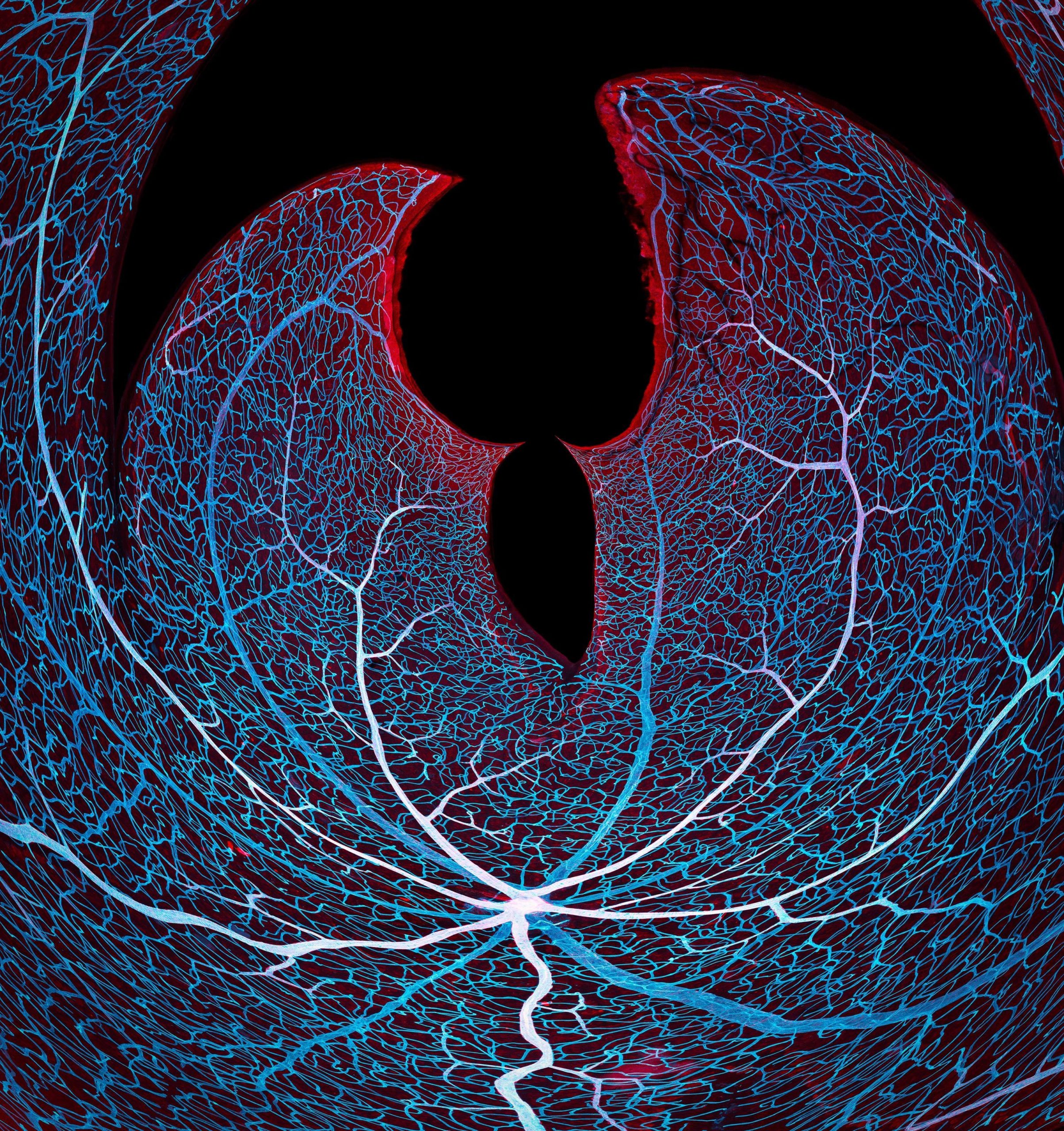
This very trippy image shows the vasculature of a mouse retina. Shown at 20-times magnification, it was taken by Jason Kirk and Carlos P. Flores Suarez from Baylor College of Medicine in Texas.
Breast organoid
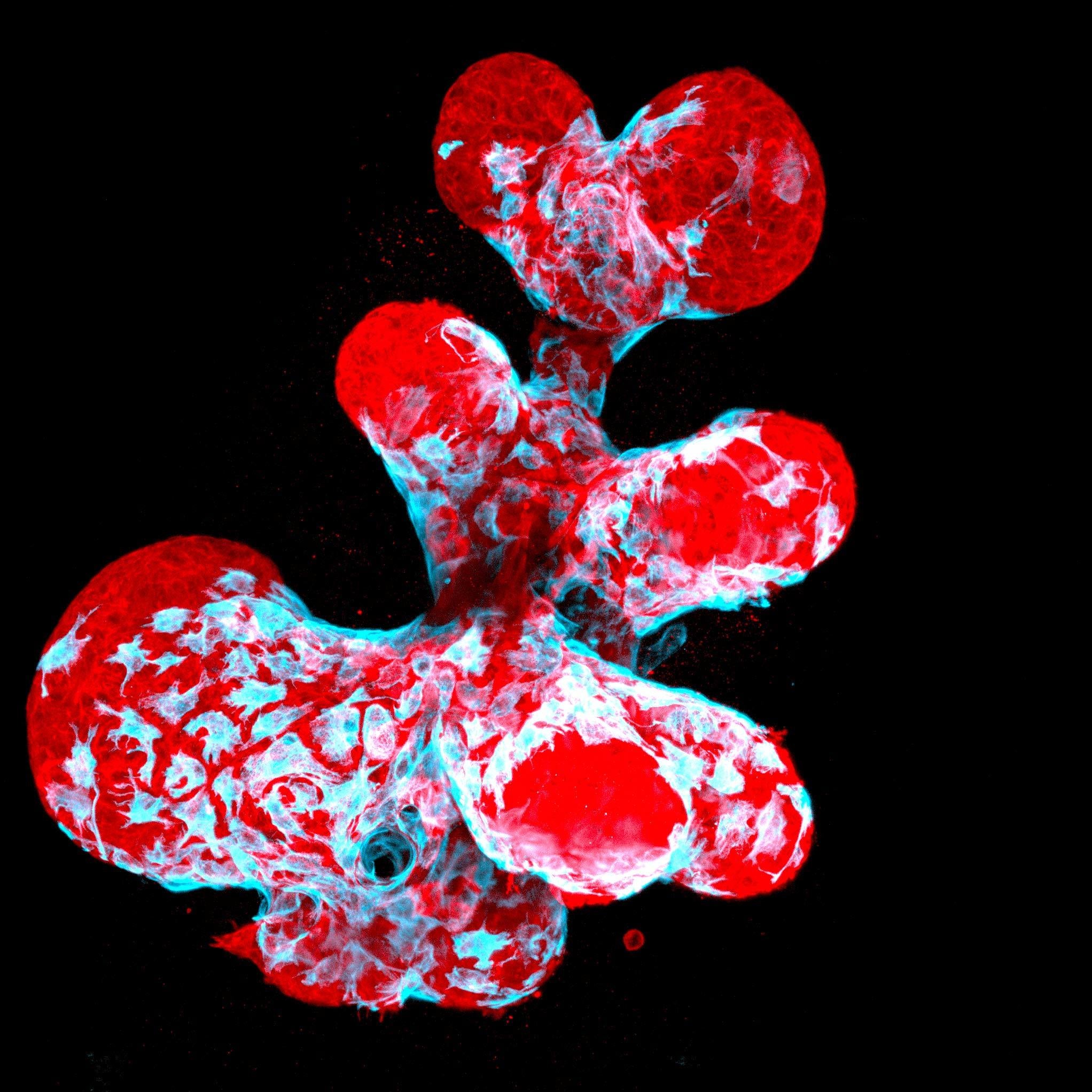
A breast organoid (a cultured and miniaturized version of the real thing) showing contractile myoepithelial cells (blue) crawling on secretory breast cells (red). Jakub Sumbal from Masaryk University in the Czech Republic was awarded 12th place for this image.
Pollen on fabric
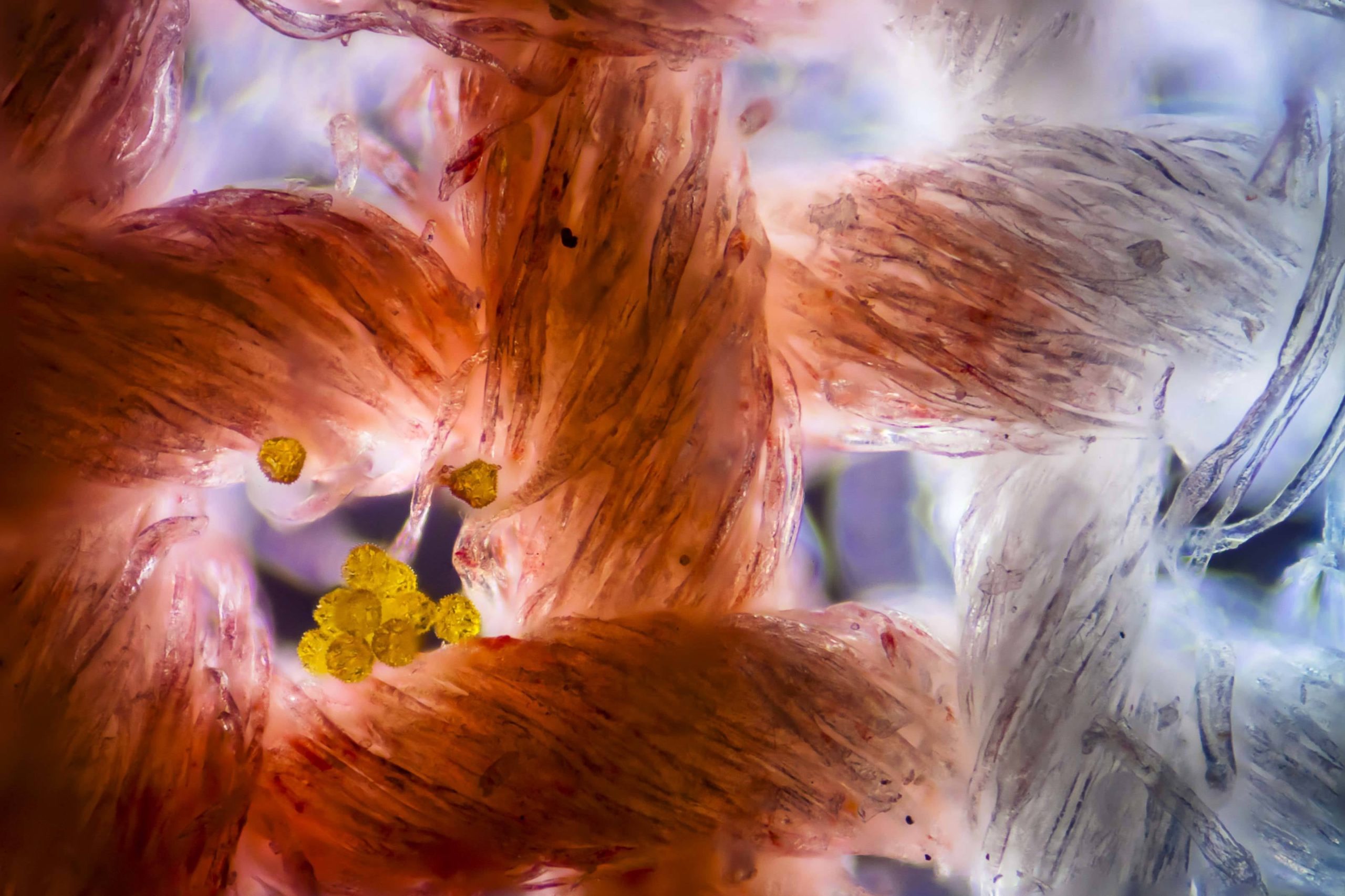
Felice Placenti of Italy took this image of cotton fabric with pollen grains attached.
Snowflake
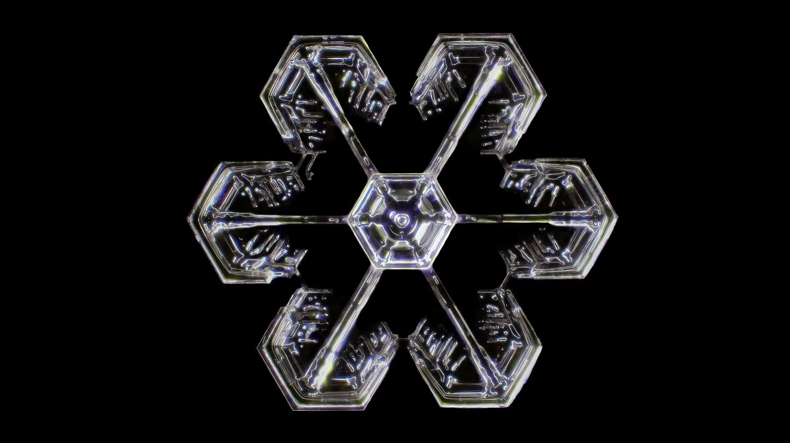
A gorgeous and almost mechanical-looking snowflake, as imaged by Joern N. Hopke from Waban, Massachusetts.
Microscopic algae
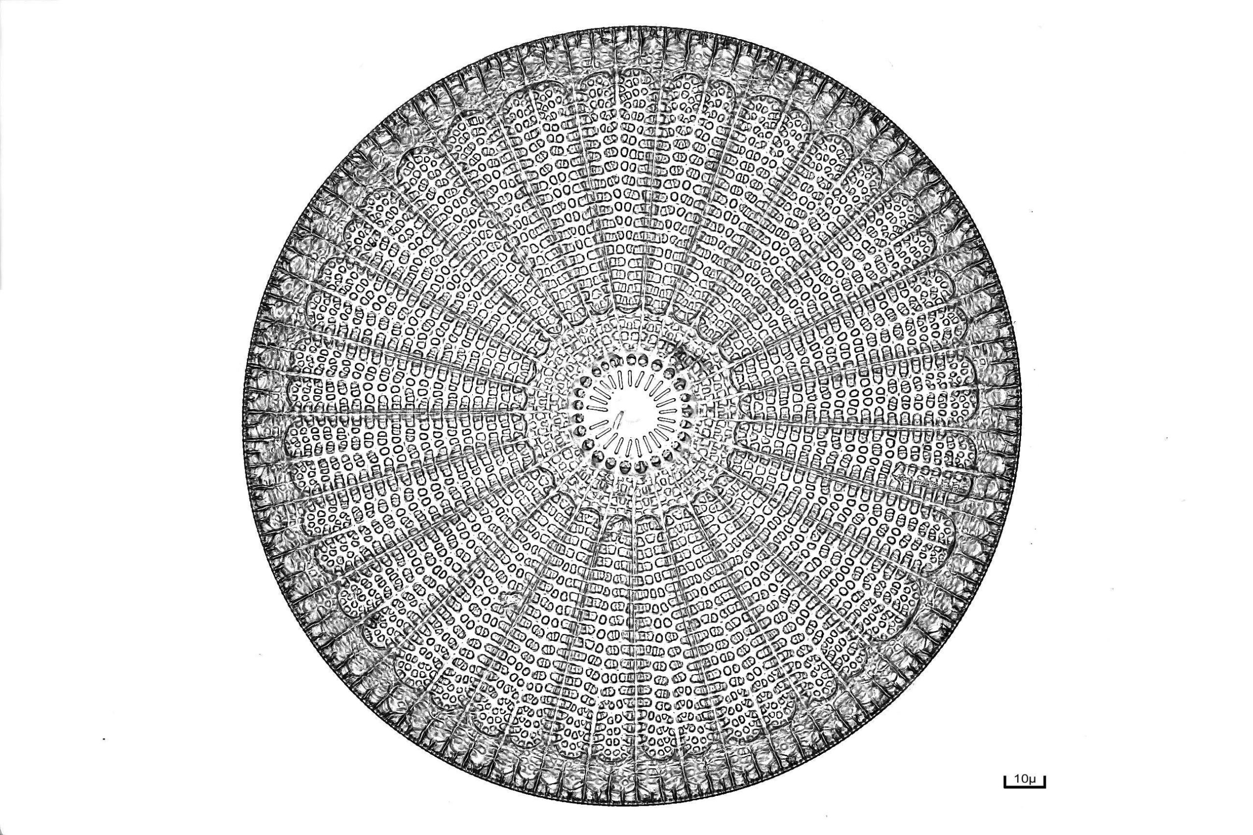
The Arachnoidiscus diatom is a single-celled organism that lives on tropical seawoods. Diatoms are famous for their symmetrical shapes, this one being an excellent example.
Cyanobacterial strands
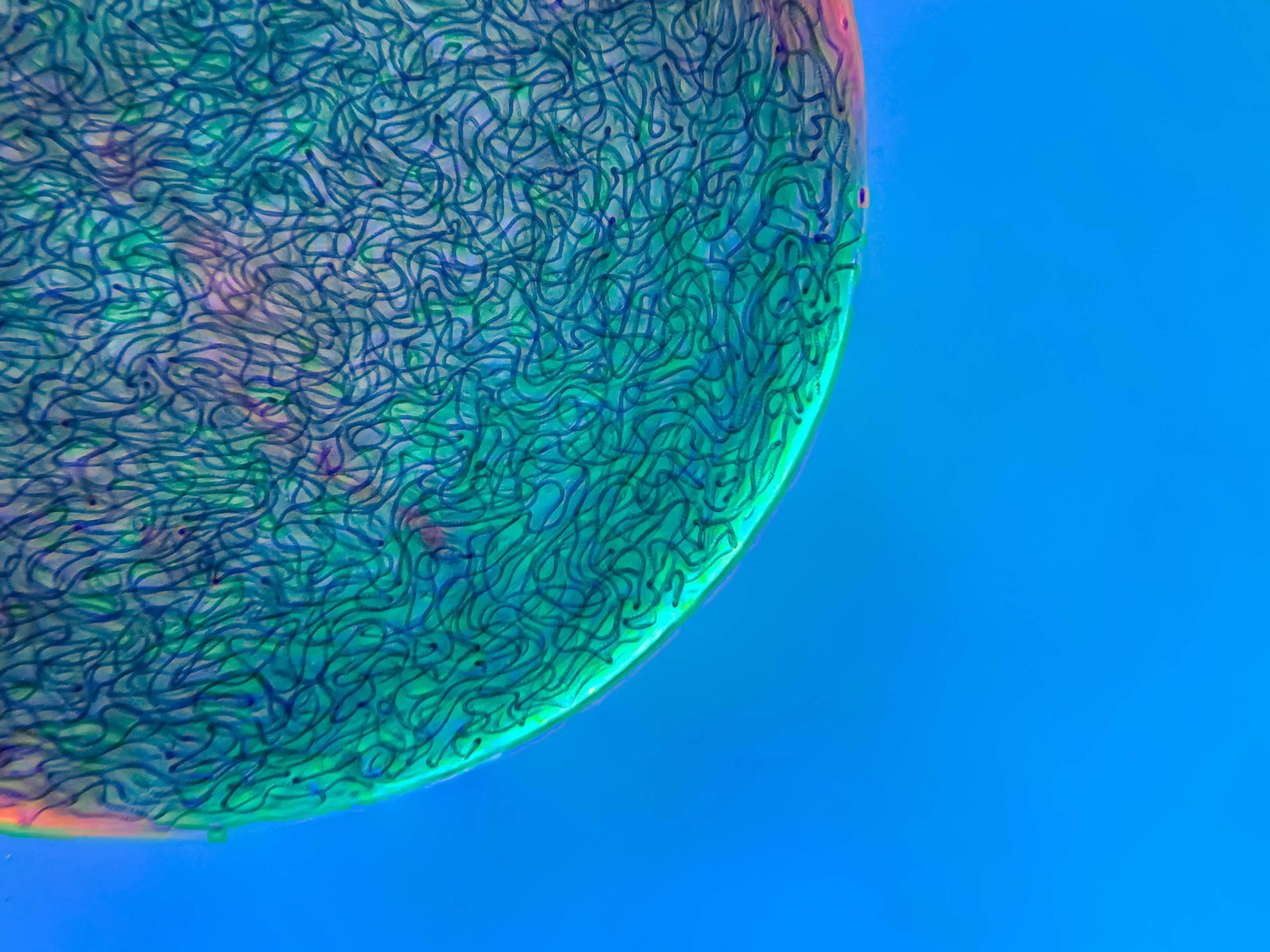
Martin Kaae Kristiansen of Denmark captured this image, showing filamentous strands of the Nostoc cyanobacteria.
Slime mould
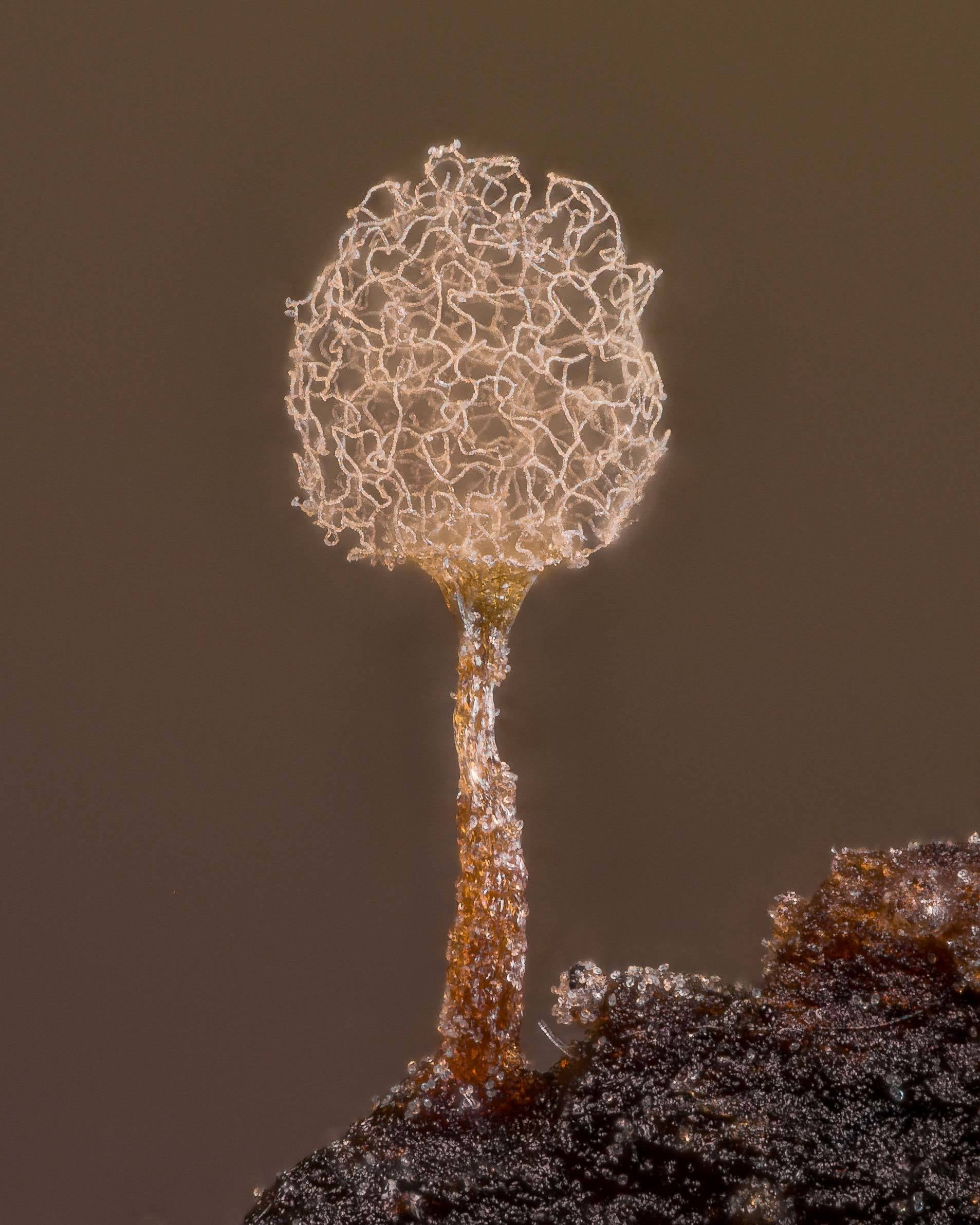
Alison Pollack of San Anselmo, California, captured this remarkable image of Arcyria pomiformis, a species of slime mould.