Pediatric cardiac surgery is one of the most demanding medical specialties. It needs to deal with delicate structures – the heart of the newborn child can weigh only 20 grams, fitting in the palm of your hand.
3D printing is rapidly changing how these surgeries can be carried out.
When the heart has only one abnormality, or a relatively common one, most doctors can rely on their experience or CT and MRI scans. But when the heart in question is tiny, the disease is rare, or is atypical in any way, even sub-millimeters in measurement can make a life-or-death difference.
This is a story of Kordian, a 3-week old infant from Poland with a potentially fatal condition known as interrupted aortic arch.
Dr Jarosław Meyer-Szary MD, from the Department of Pediatric Cardiology and Congenital Heart Defect at the University Clinical Center in Gdańsk, Poland made the decision to use a to-scale 3D printed model of Kordian’s heart as a support.
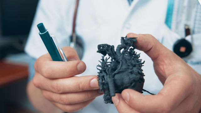
One of the benefits of 3D printing, used by cardiologists with increasing popularity, is the ability to print and see the abnormality close-up, before the surgery – even performing a “practice” surgery on it.
For full-sized adult hearts, finding a 3D printer capable of this is pretty simple. For Kordian’s surgery, every tiny little vein and artery that surrounds the heart needed to be recreated with perfect accuracy and strength. So SLS (selective laser sintering) 3D technology was used. Sinterit Lisa, a desktop-size selective laser sintering 3D printer, was used in this case.
Kordian is now 18 months old.