Mouse brains! Bat embryos! Hungry algae! Today, Olympus unveiled the winners of the tenth annual BioScapes photography competition, which showcases the best photography captured through light microscopes. The top 10 were chosen from a mind-boggling 2,100 entries.
What’s really cool about BioScapes is how drastically imaging technology has improved since the competition began. Honorable Mention
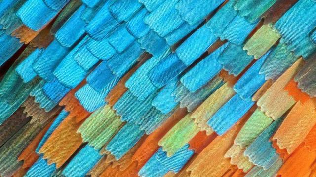
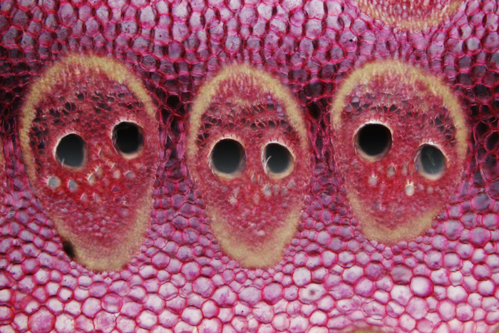
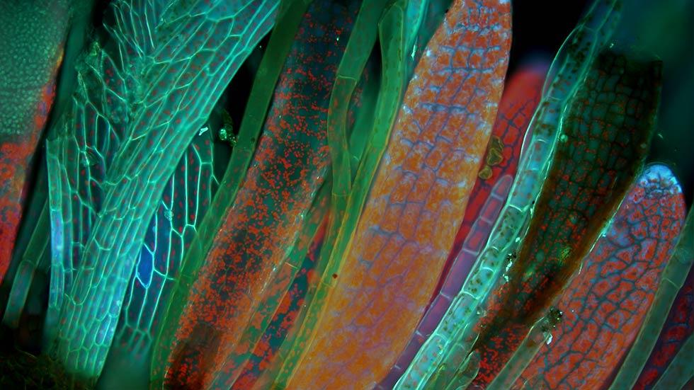
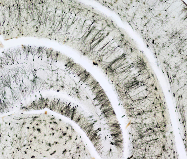
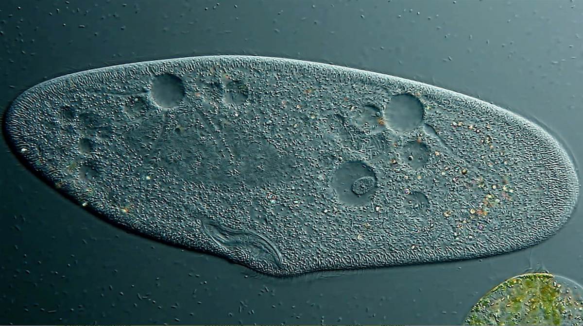
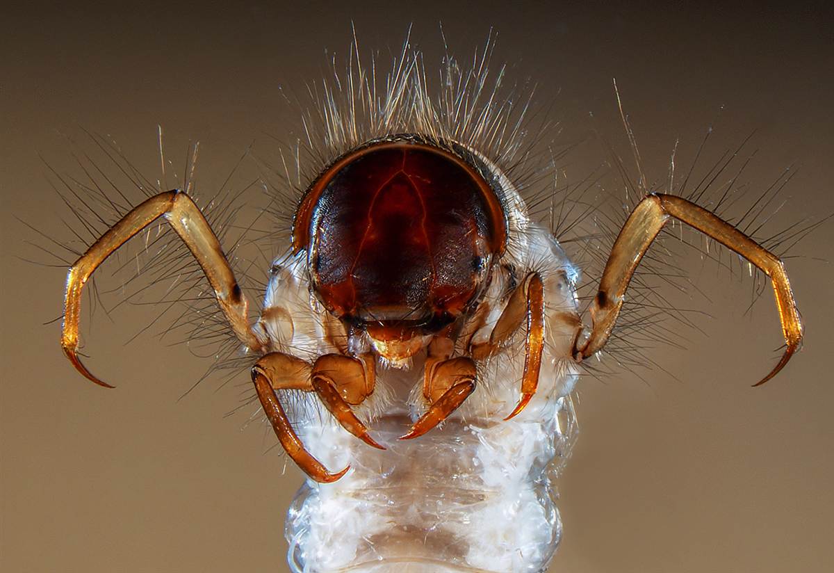
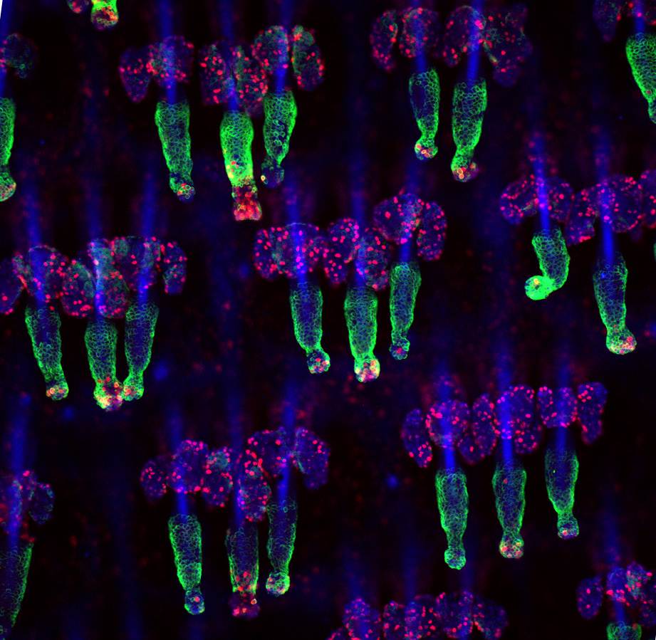
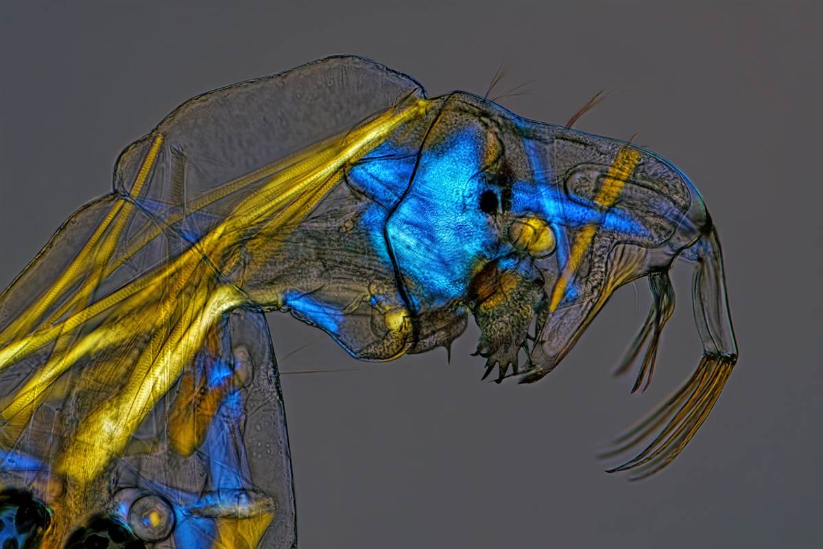
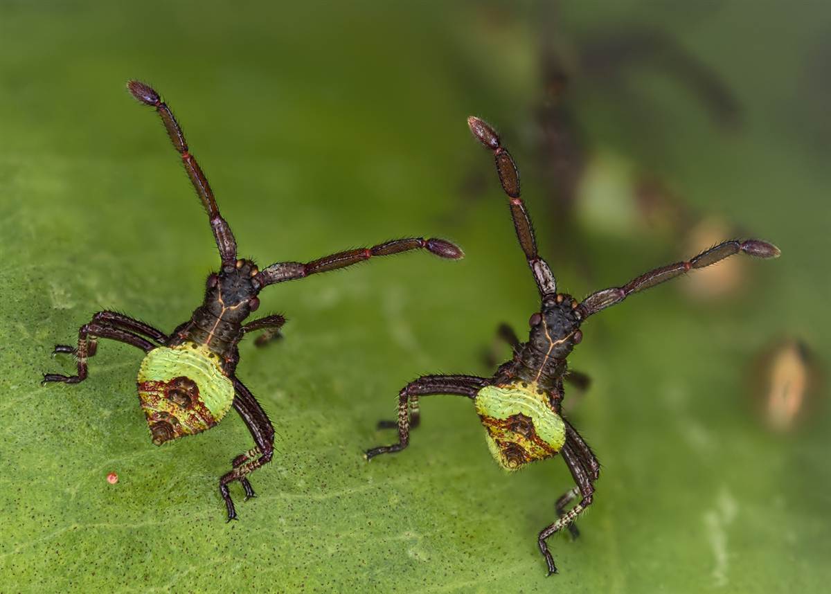
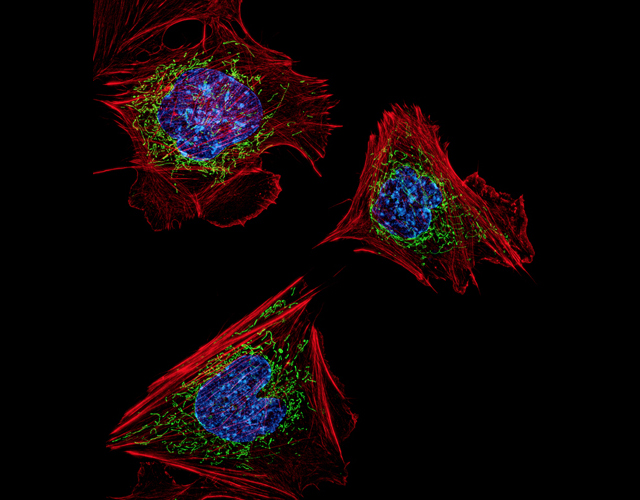
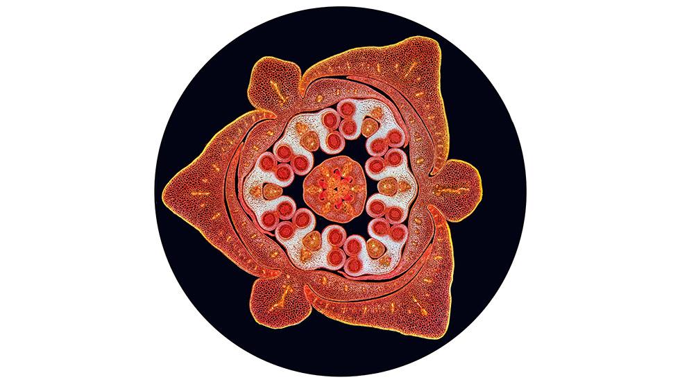
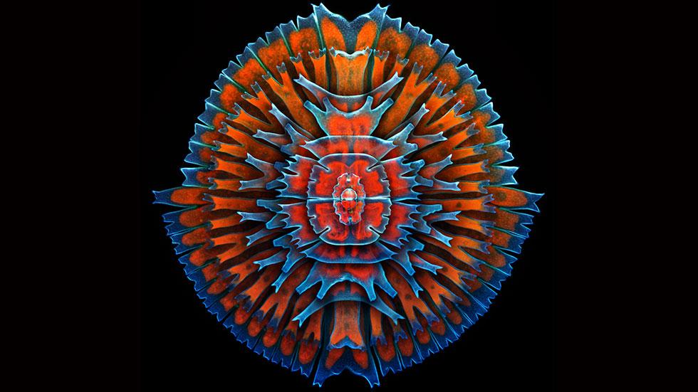
Charles Krebs, who won Bioscapes’ seventh place prize, also received an honorable mention for this lovely, Georgia O’Keeffe-esque image of a dissected Camellia bud — it shows immature anthers and filaments against the unopened flower petals. Norfolk’s Dr. David Maitland Feltwell captured this image of the Cocoa nut palm (Cocos comosa) stem with xylem vessel “eyes” in vascular bundle “faces.” University of Wrocław’s Magdalena Turzańska University of Wrocław captured a beautiful image of Lepidozia reptans, aka the pinnately branched leafy liverwort — a common type of buttercup! Stanford scientist Ahmad Salehi, MD, used brightfield micsoscopy to reveal the structure of the brain of a mouse — specifically, the hippocampal region. A paramecium — a protozoa used in science classrooms and labs as an example of a basic organism — is the subject of this image by Ralph Grimm of Jimboomba, Australia. These little dudes live in fresh water. They actually use water to propel themselves, too, by sucking it in and out of two vacuoles at each end of its body. The caddisfly — a common type of moth-like fly that nests in water and soil — builds its own protective case, often out of silk and bits of sand or soil. This image by Fabrice Parais of Normandie, France, shows a larvae with a translucent case. These little dongles are actually mouse tails, which have been stained to illuminate the hair follicle stem cells. The image was captured using confocal imaging by NYC scientist Yaron Fuchs at the Howard Hughes Medical Institute. Glassworms — aka phantom midge larva (Chaoborus) — are found in lakes all over the world. But, as its name suggests, it’s usually hard to spot given its translucent skin. Washington scientist Charles Krebs used specialised illumination to reveal the the structure of these nearly invisible larvae. Basel scientist Kurt Wirz wins for the most heartwarming entry: His image, entitled Brother Bugs, shows two-hour-old squashbugs enjoying their first moments together. Dr. Dylan Burnette, from the National Institutes of Health, illuminated the embryonic fibroblasts of mice to show their basic structure — including actin filaments (red), mitochondria (green) and DNA (blue). UK scientist Spike Walker used darkfield illumination to capture a section of the Lily flower bud, which is just as exotic on the microscopic level as on the visible scale. Dr. Igor Siwanowicz, a neurobiologist at the Howard Hughes Medical Institute in Virginia, shot this single-cell fresh water algae (desmids) — magnified by 400x, of course. An Oxford scientist named Dorit Hockman took this shot of a bat embryo using stereo microscopy. It shows an unborn black mastiff bat Molossus rufus, wrapped in alien-esque white wings. Siwanowicz took home first as well as third place — his winning image shows the gaping maw of an aquatic carnivorous plant, the humped bladderwort Utricularia gibba. “The plant feeds on microinvertebrates, and sucks them in within a millisecond after they touch the trigger hairs growing from the center of the plant’s dome-shaped trap,” explains the Howard Hughes Medical Institute. Those neon circles inside? Those are single-cell organisms that have just been devoured. The lead image by David Millard of Austin, Texas, shows the body scales of a great purple hairstreak butterfly (Atlides halesus). Check out the rest of the images over at the BioScapes page.
Honorable Mention
Honorable Mention
Honorable Mention
Tenth Place
Ninth Place
Eight Place
Seventh Place
Sixth Place
Fifth Place
Fourth Place
Third Place
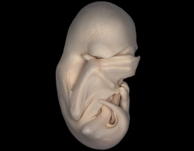
Second Place
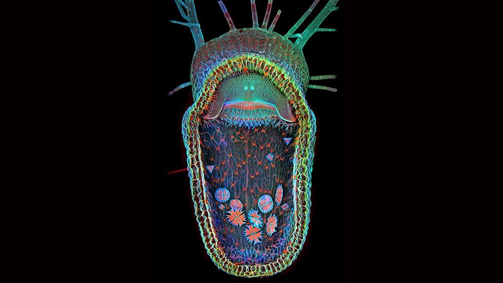
First Place