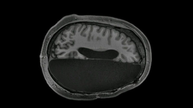
In rare cases, patients suffering from severe epilepsy undergo an operation in which an entire brain hemisphere is removed. New research shows the startling degree to which the remaining hemisphere is able to pick up the slack.
The human brain has an amazing ability to reconfigure itself following a dramatic loss of function, a phenomenon that has been known for decades. New research published this week in Cell Reports adds to this body of knowledge, with researchers examining six adult patients who underwent hemispherectomies as children. The patients exhibited surprisingly strong neural connectivity between different parts of their remaining brain, the study found.
The new work, co-authored by neuroscientist Dorit Kliemann from the California Institute of Technology, provides insights into how the brain reorganizes itself after a radical loss of brain material. The research is evidence of “compensated cognition,” the scientists write.
“The people with hemispherectomies that we studied were remarkably high functioning. They have intact language skills; when I put them in the scanner we made small talk, just like the hundreds of other individuals I have scanned,” said Kliemann in a statement. “You can almost forget their condition when you meet them for the first time. When I sit in front of the computer and see these MRI images showing only half a brain, I still marvel that the images are coming from the same human being who I just saw talking and walking and who has chosen to devote his or her time to research.”
Indeed, individuals with hemispherectomies can have quite good cognitive health despite the severe nature of the procedure. The new research was an effort to learn more about the neurological processes responsible.
The participants recruited for the study included four men and two women, all now in their 20s and 30s, who had hemispherectomies when they were between the ages of 11 months and 11 years old. An additional six participants with intact brains made up the control group.
Using an fMRI machine, the scientists looked for signals associated with spontaneous brain activity while the participants were in a relaxed, resting state. The researchers focused on the brain regions known to be responsible for vision, movement, emotion, and higher thinking. This data was then compared to a pre-existing database, the Brain Genomics Superstruct Project, containing the brain scans of 1,842 normally functioning individuals.
It wouldn’t have been unreasonable to expect the scans to show radically rearranged brain networks, since a single hemisphere is now carrying out the work typically done by two. But that’s not what they saw.
Surprisingly, the brain activity of the hemispherectomy patients appeared largely typical, showing the same resting-state networks one would see in a normal brain. But there was one major exception: The single-hemisphere brains showed greater-than-normal connectivity between networks than the control brains did.
“These results… suggest that between-network interactions may characterise functional reorganization in hemispherectomy,” the authors write in the study. It remains an open question as to how the increased connectivity might assist—or even impair—functioning among hemispherectomy subjects, such as their sociability, motor skills, and decision-making. Future research “will need to investigate the behavioural correlates” of these connections, according to the paper.
The new work is preliminary, and looking ahead, the team would like to expand their research to verify these results among a larger sample size, among other improvements to their experimental design.
The hemispherectomies were performed when the study participants were children, which likely made it easier for their brains to adapt to such a dramatic alteration. Insights from this research could shed new light on how the brain reorganizes itself following a traumatic injury and loss of neurological function.
“As remarkable as it is that there are individuals who can live with half a brain, sometimes a very small brain lesion like a stroke or a traumatic brain injury like a bicycle accident or a tumour can have devastating effects,” said Kliemann. “We’re trying to understand the principles of brain reorganization that can lead to compensation. Maybe down the line, that work can inform targeted intervention strategies and different outcome scenarios to help more people with brain injuries.”
There’s still so much to learn about the human brain—which is at once incredibly fragile, yet also super resilient.