A new microscope developed at the Marine Biological Laboratory in Woods Hole, Massachusetts is allowing scientists track the position and orientation of individual molecules in living cells. It has the potential to reveal unknown aspects of molecular behaviour, including those that turn cells into agents of disease.
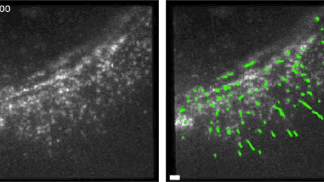
Left: Fluorescent molecules move and assemble inside a living human skin cell. Right: The green strips indicate the orientation of the individual molecules. (Credit: Shalin Mehta and Tomomi Tani)
Dubbed the “instantaneous fluorescence polarization” microscope, this new tool is being used to understand how tiny molecules move and assemble inside live cells, including human skin cells. It reveals how individual molecules — which measure just a billionth of a metre across — wiggle around a live cell, bind together to form larger cellular structures and drive a cell’s biological functions.
Left: Fluorescent particles move along actin filaments, which allow cells to contract, in a human skin cell. Right: Pink lines show the orientation of the actin filaments. (Credit: Shalin Mehta and Tomomi Tani).
As described in a new paper in Proceedings of the National Academy of Sciences, scientists were able to determine the orientation of single molecules, and watch how an assembly of molecules came together to form a higher-order structure.
“Cells rely on a wide variety of molecular arrangements for their function,” lead author Shalin Mehta of the MBL and the University of Chicago told Gizmodo. “For example, muscles contract along a specific orientation because of the alignment of molecules. This new microscope and algorithms let scientists see how individual molecules align and interact with each other in live cells, even though these assemblies are much finer than the resolution limit of the light microscope.”
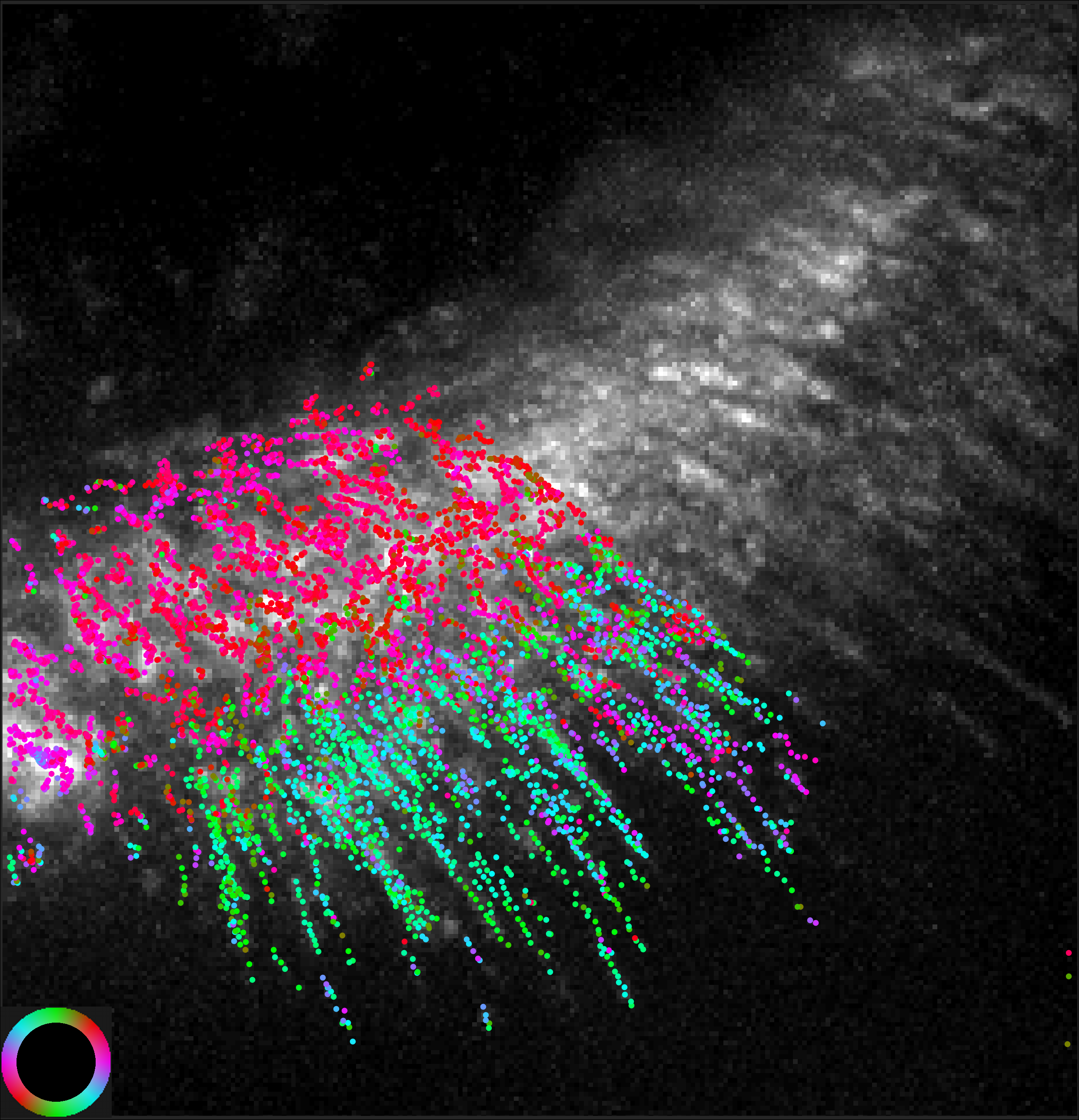
The orientation of actin filaments (colour-coded) is overlaid on an image of fluorescence intensity. (Credit: Shalin Mehta and Tomomi Tani)
Meha and his team were able to catch a glimpse of the microscopic particles by using polarised light, a property of light that’s not visible to the human eye. After labelling DNA and actin molecules with fluorescence, the researchers tracked the movements of these tiny particles in living human skin cells.
This microscope will improve our understanding of cellular function, and potentially explain why cells sometimes go haywire, such as when they turn cancerous.