Image Cache: Every year, Nikon’s Small World Competition rounds up the best microscopic images from scientists around the world. The results are strange and beautiful — take a look at them here.
The competition brings what scientists see through their microscopes on a daily to the wider world. The winning entries are always impressive, and this year’s are no different.
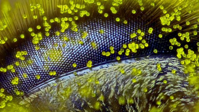
Taking first place this time round is Ralph Grimm from Queensland, Australia, who captured this image of the eye of a honey bee covered in dandelion pollen. Magnified 120 times, it reveals the microscopic complexity of the optical system of the insect.
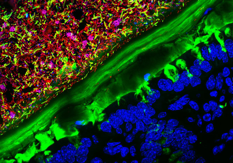
In second place, this image by Kristen Earle, Gabriel Billings, KC Huang & Justin Sonnenburg shows a section of mouse colon colonised with human microbiota. You can see the bacteria in red, the colon tissue in blue and the layer of mucus that separates them in green.
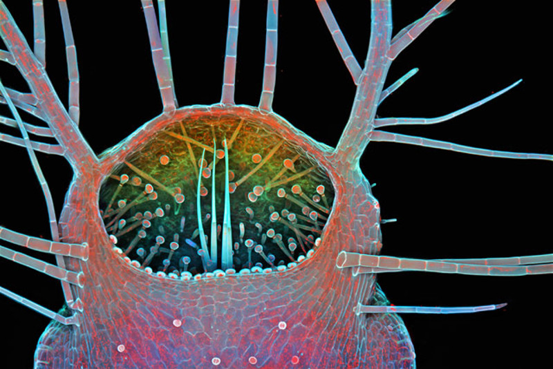
This alien-looking image is in fact the intake of a humped bladderwort — a kind of freshwater carnivorous plant — magnified by 100 times. It was captured by Dr. Igor Siwanowicz from the Howard Hughes Medical Institute, and snatched third place.
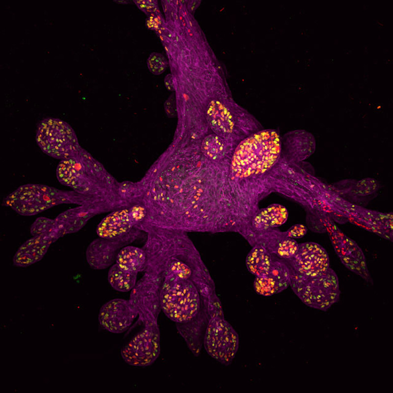
In fourth place came this image of part of a lab-grown human mammary gland. It was taken by Daniel H. Miller & Ethan S. Sokol at the Whitehead Institute for Biomedical Research.
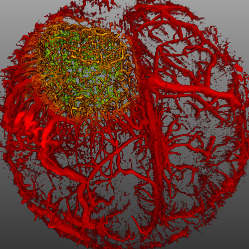
And finally, in fifth place was this live image of the blood flow in a mouse brain, captured using optical frequency domain imaging. It was captured by Dr. Giorgio Seano & Dr. Rakesh J. Jain from Harvard Medical School.
If you like what you’ve seen, you can see the rest of the top 20 images, along with other honorable mentions, over on the Nikon Small World website. Go check it out, it’s worth a look.
All images by the photographer and Nikon Small World