What does a tear look like at the microscopic level? It’s more interesting — and beautiful — than you might expect.
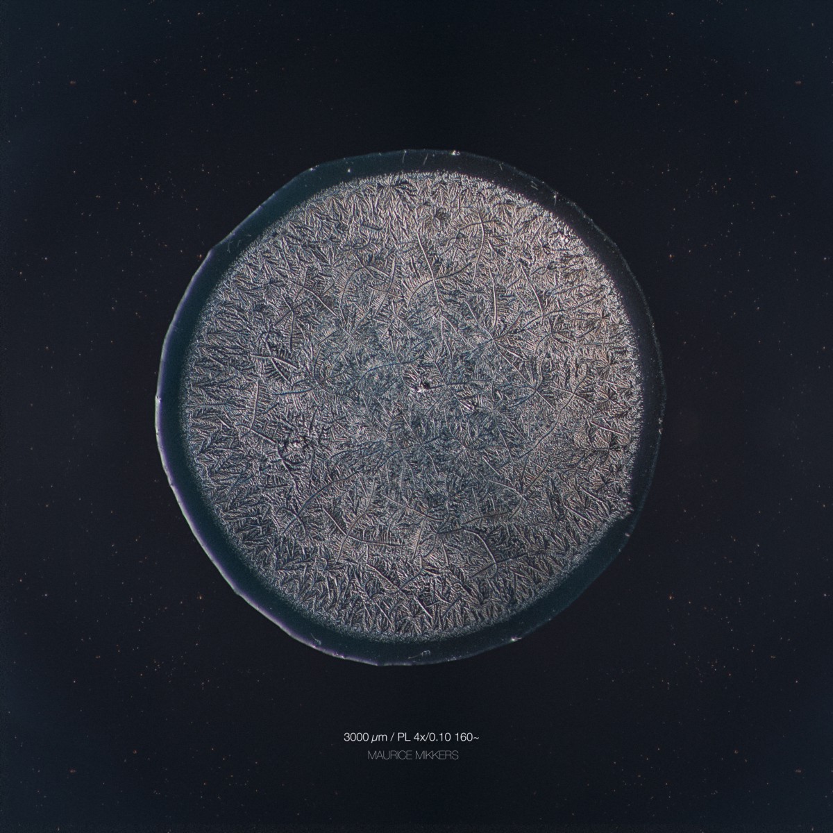
Dutch photographer Maurice Mikkers recently captured a few really amazing close-up images of the most precious human body fluid: tears. The project is a lovely illustration of how our emotions, from pain to joy, are reflected in the tears that protect our eyes. Mikkers — who is a licensed Medical Laboratory Analyst by the way — began the project by considering the scientific fact that there are three kind of tears: basal tears, reflex tears, and emotional tears, asking a simple question:
Is there any difference? Science says that every tear has a different viscosity and composition. All tears contain a variety of biological substances including oils, antibodies and enzymes suspended in salt water. But how does this relate to the “real world”. Because of this I decided to start an evening of experimenting with my close friends. I asked them to cut unions, eat hot peppers, look in to a fan or cry because of sadness or happiness. To see if there was a resemblance or difference in the structure of forming tears, I took images of every tear drop under the microscope.
Mikkers captured the tears of his friends with a micropipet, and dispensed the teardrops onto a microscopic slide. After a while the different fluids started to crystalize and settle, so it became possible to take images and begin the comparison:
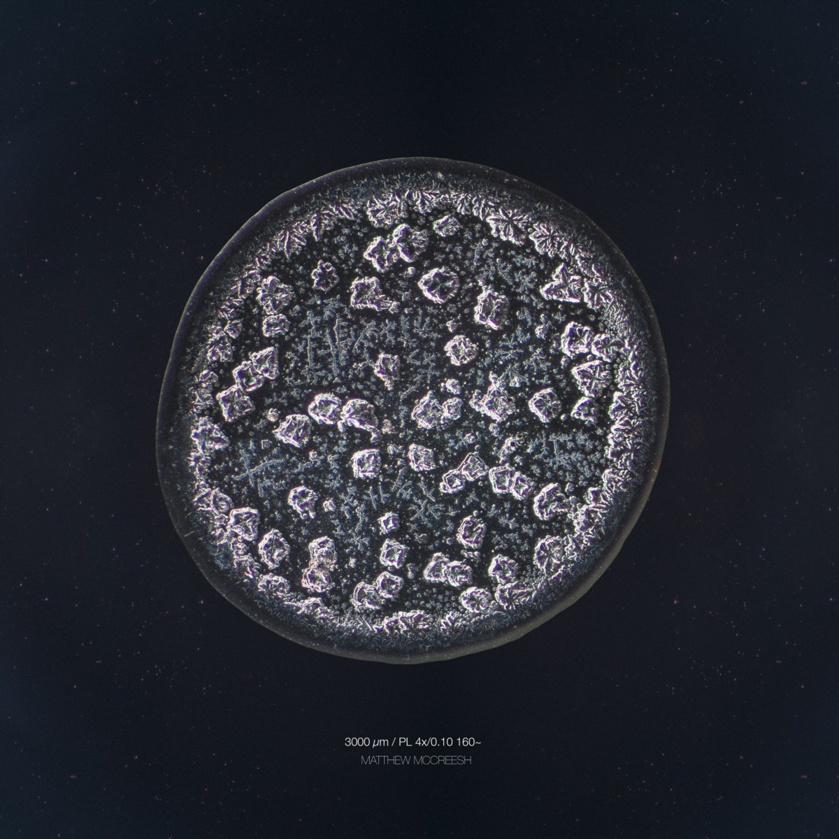
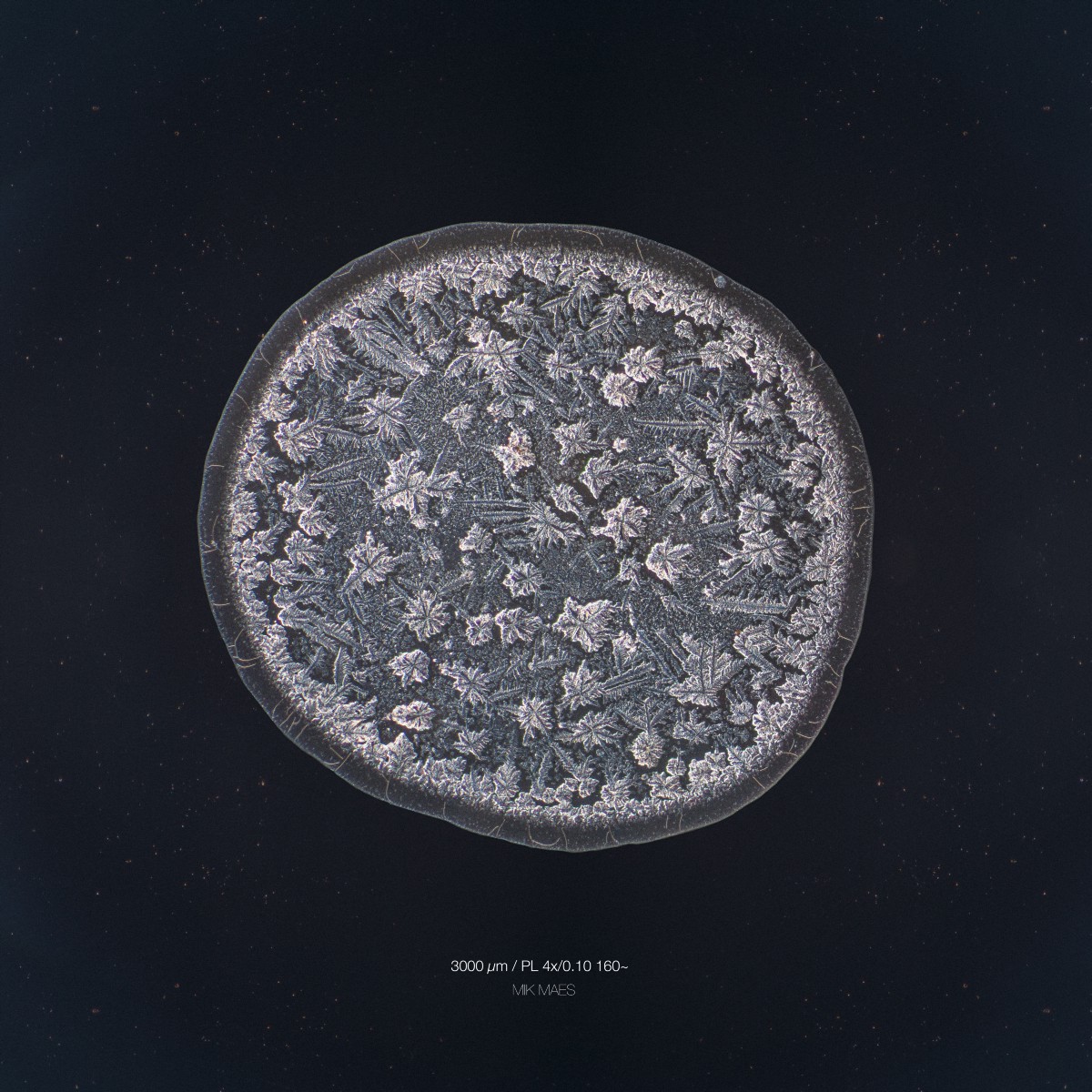
The structures seen under the microscope and in the images are largely crystallised salt, the circumstances under which the tear dries can lead to radically dissimilar shapes and formations, so two psychic tears with the exact same chemical makeup can look very different up close.
Just look at the following set of micrographs in order to see the difference by yourself.
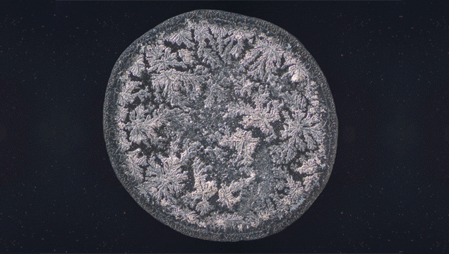
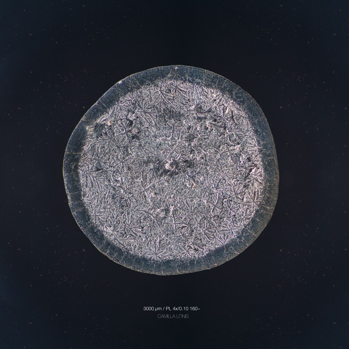
Reflex tears: harvested after cutting white onions

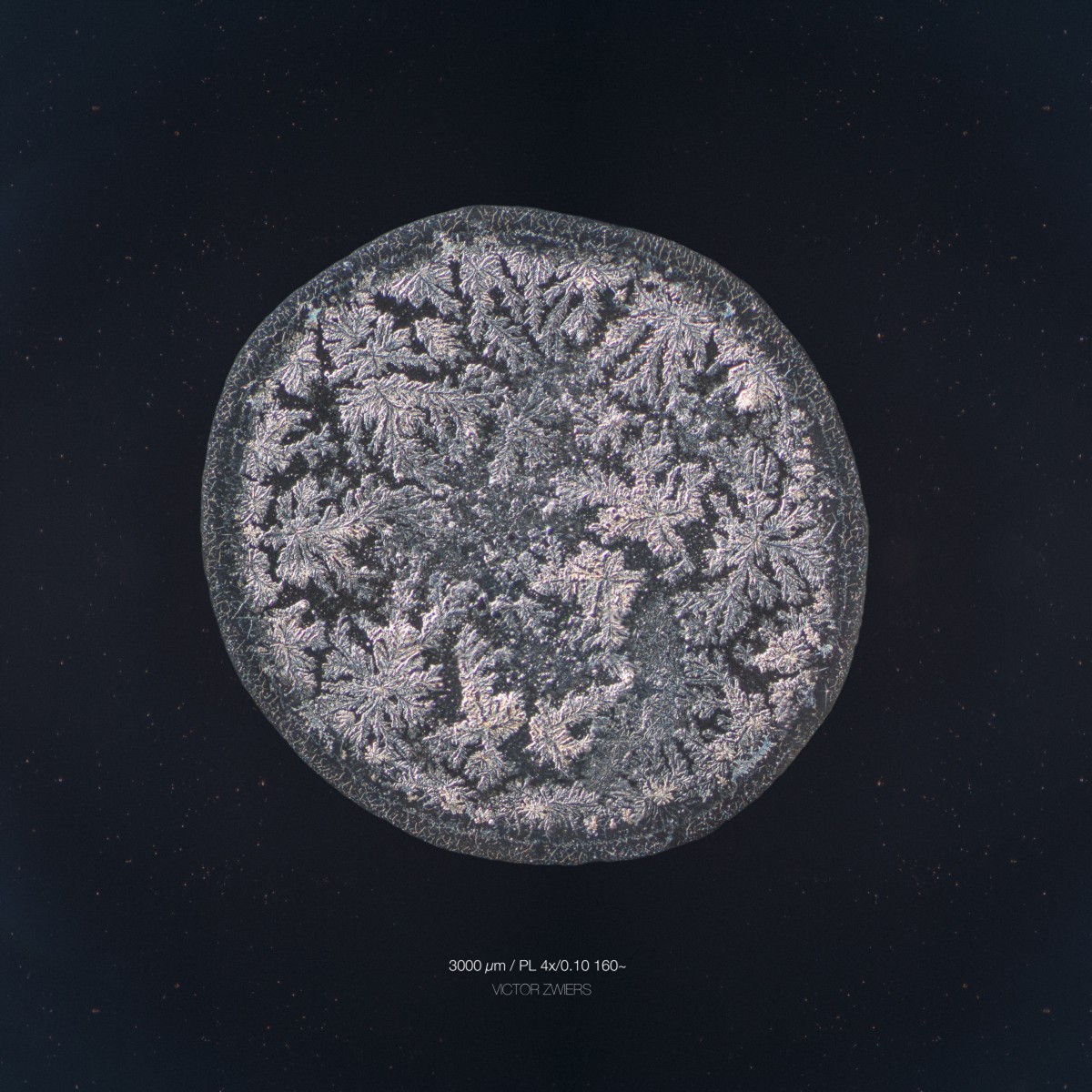
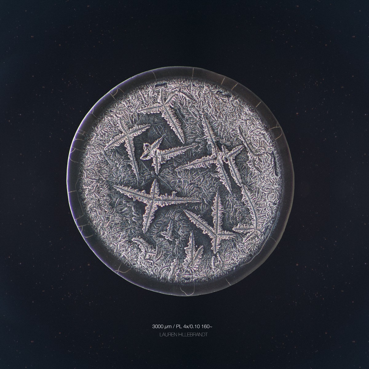
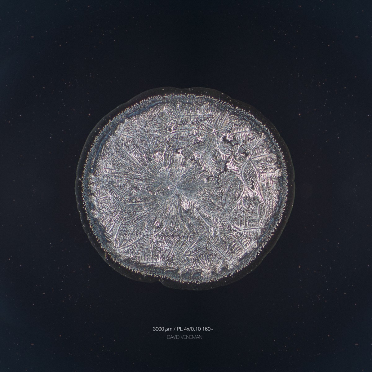
Psychic tears: harvested after a forced emotional response
Reflex tears: harvested after cutting white unions and eating a red pepper
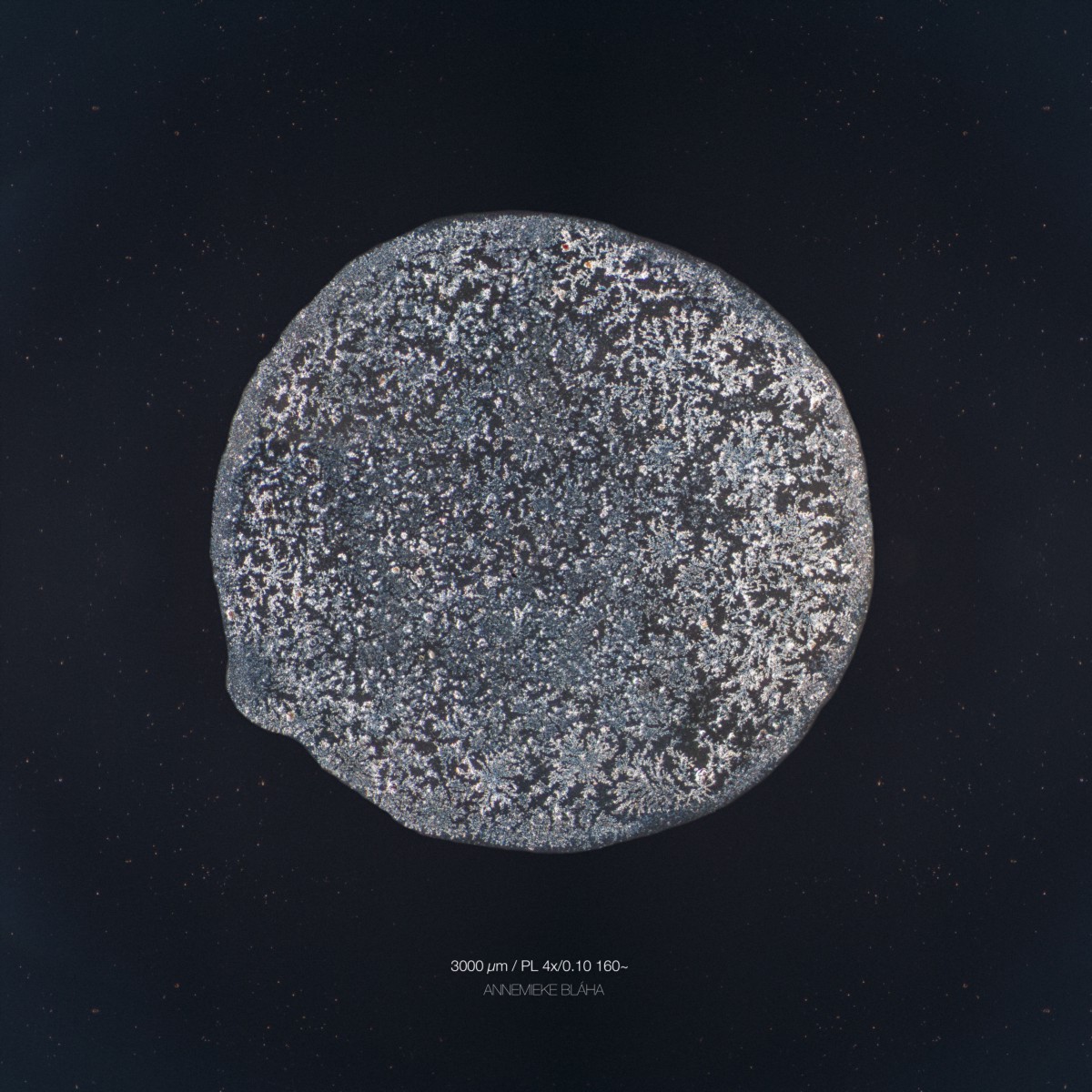
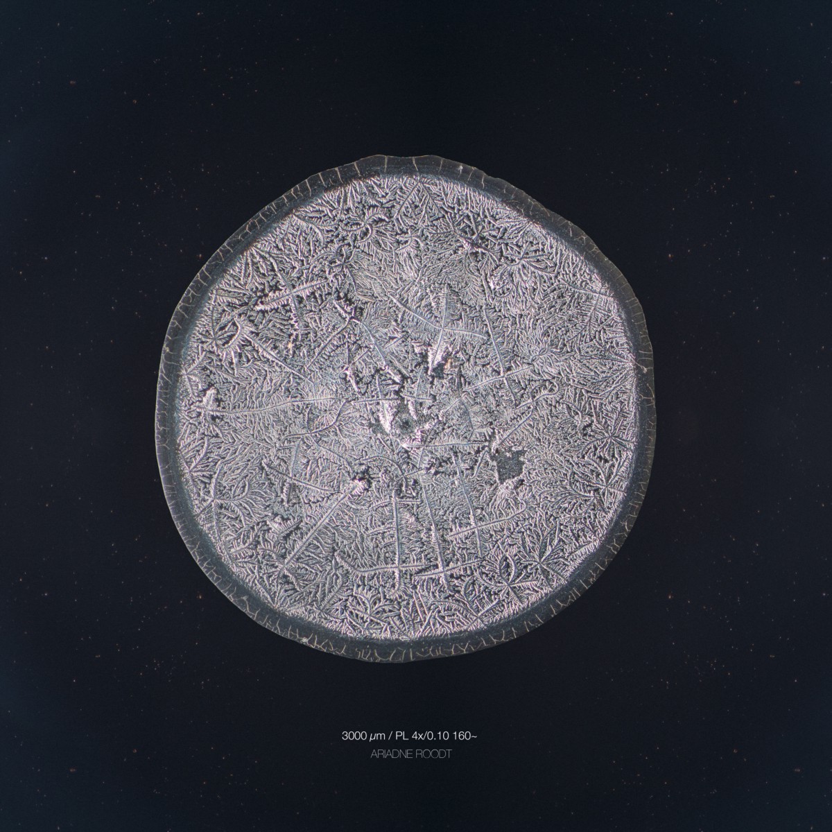
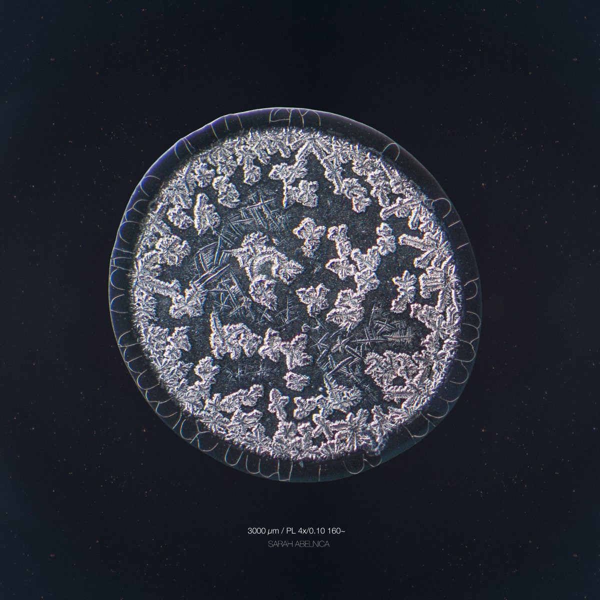
Basal tears: harvested after looking in to a fan for a few minutes
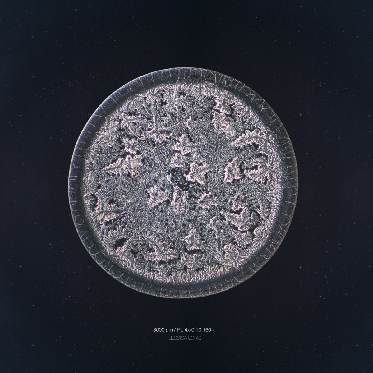
Reflex tear: harvested after eating a red pepper
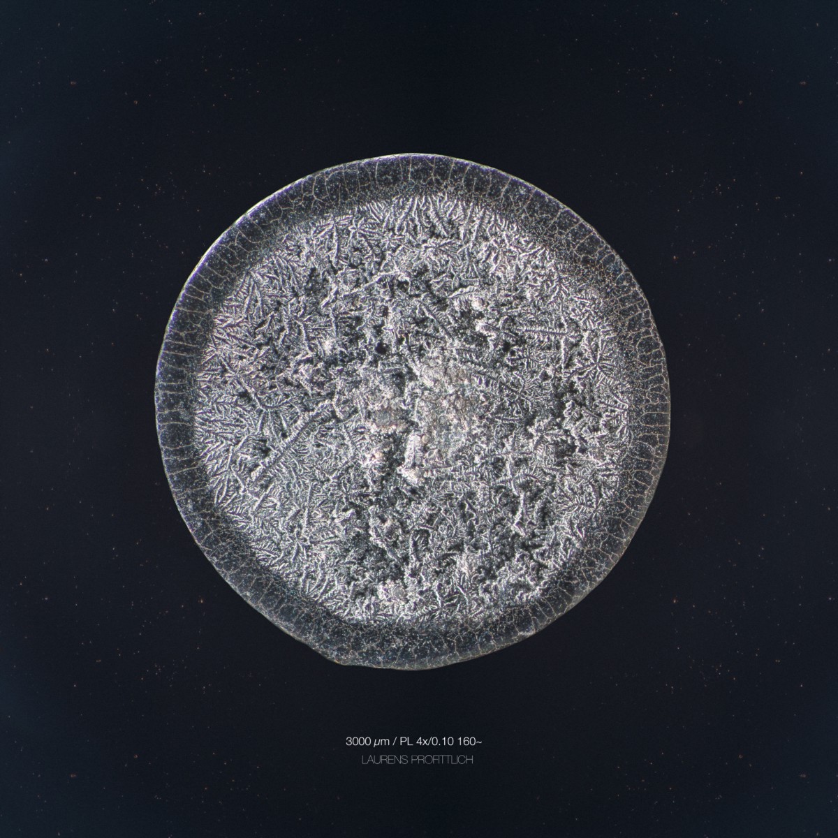
Reflex tear: harvested after eating a mini red chilli pepper
Reflex tear: harvested after crying from high dosed menthol oil on eyelid
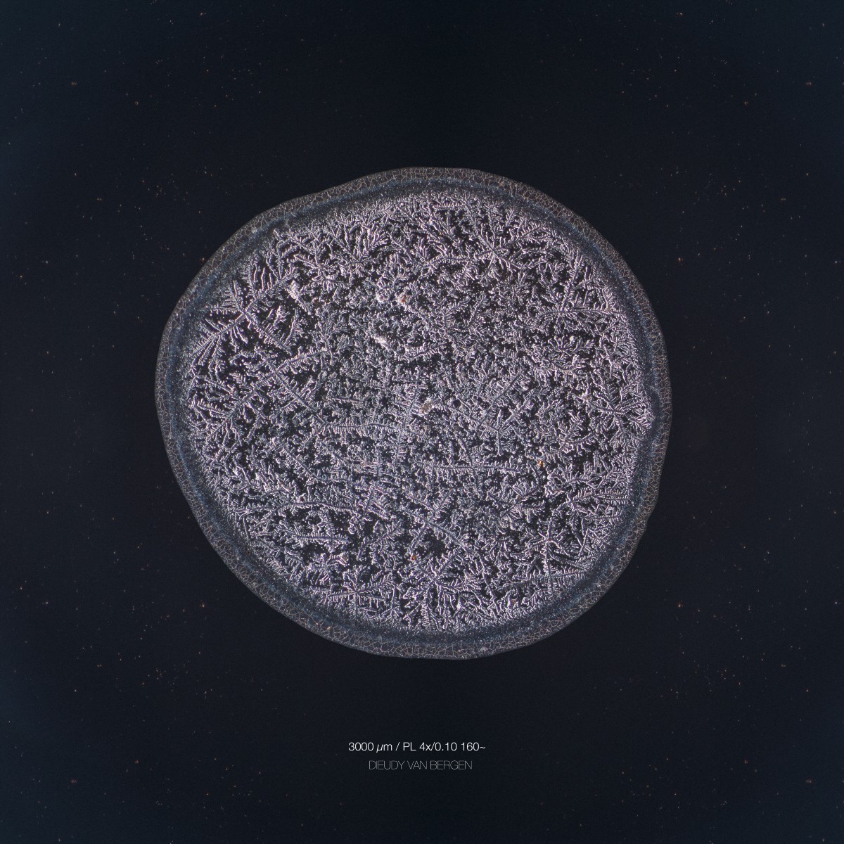
Reflex tear: harvested after cutting white unions, putting pepper in the eyes, snorting water and eating a red pepper