The magnetic resonance imaging (MRI) machines in hospitals are great at creating pictures of the human body. For decades, scientists have hoped that the same technology could be used to examine much smaller things, like individual molecules. Now, a team from Canada and the United States has revealed a new, high-resolution MRI method with resolutions down to two nanometres, the width of a DNA strand.
The researchers combined a special kind of magnetic field generator and specifically engineered laser pulses to detect the properties of atomic nuclei and control those properties during the imaging. It’s as if they combined the best dye, microscope, and tweezers to make incredible images of proton behaviour at a two-nanometre spatial resolution. This advancement could be especially useful for viewing and characterising molecules in biological samples and other microscopic systems.
“Methods like these could even be revolutionary for the understanding of molecular dynamics,” Elizabeth Donley, scientist in the Atomic Devices & Instrumentation Group at the National Institute of Standards and Technology, who was not involved in the research, told Gizmodo in an email.
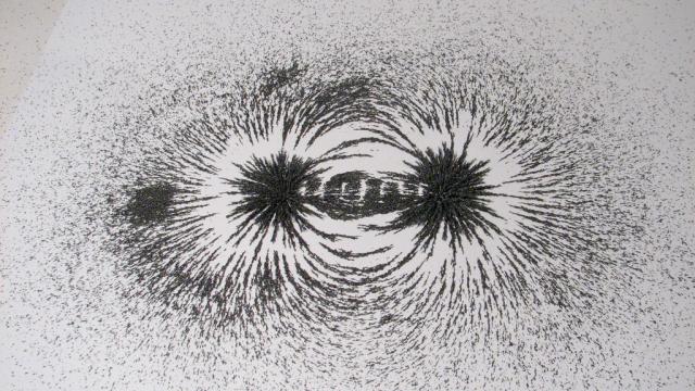
MRIs make their images using nuclear magnetic resonance, or NMR. The nuclei of some atoms absorb and re-emit radio waves in a strong magnetic field. Understanding the characteristics of the emitted radio waves, like their wavelengths, provides information about the complexities of the electric fields around the atoms. On larger scales, this can make some pretty crazy pictures. On smaller scales, it could help determine the identity and structure of molecules in fine detail. Scientists want to bring MRI to the smallest possible length scales to better understand tiny things like proteins.
The true challenge is sensing a fundamental property of particles that gives rise to magnetic fields called the “spin” on the smallest scales, according to the paper published by a team led by Raffi Budakian, professor at the University of Waterloo in Canada, in the journal Physical Review X.
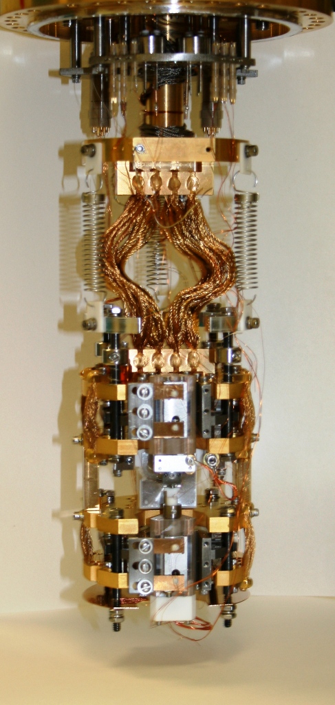
Raffi Budakian’s team’s force microscope for making measurementsPhoto: Raffi Budakian (University of Waterloo)
So, how did the team do it? A tool called a current-focusing field gradient source (CFFGS) produces a strong magnetic field that changes quickly over short distances. This allowed the researchers to identify the radio emission frequencies of the nuclei, and where they came from, to high resolution. They also hit the samples with laser pulses, giving nanoscale control over the protons’s spins in order to separate them from the influence of the changing magnetic environment.
This isn’t the only way to image molecules at this level, but it’s one that other researchers are really excited about. It shows the true potential of MRI.
“We think the technology is maturing to the point where we can really start using MRI as a more general tool for atomic-scale characterization of materials,” Budakian told Gizmodo.
Others agreed. “The nice thing about this paper is that it really addresses some of the limitations of other nano-MRI techniques,” said Ania Bleszynski Jayich, principal investigator at the Quantum Sensing an Imaging Lab at the University of California Santa Barbara. “It’s a pretty significant step in terms of the combination of spectral and spatial resolution in the same paper.”
The imaging here was only done in one dimension, meaning along a line, but this is something all of the sources I spoke with felt wasn’t an insurmountable barrier. Budakian said that the group has already filed a patent demonstrating the imaging in all three spatial dimensions. Donley did point out that the experiment also occurred at only four degrees above absolute zero – this keeps molecules still, but means the method might not be able to capture more dynamic physics.
But applications of the method go beyond just imaging biological molecules. Sarah Li from the University of Utah thought it could be useful for optimising semiconductors or other microscopic-scale electronics. Essentially, this is a fundamentally different way of characterising things on the smallest scale.
Budakian said: “If you ask what’s I’m really excited about, it’s the opening of the door to modalities of magnetic resonance that were envisioned decades ago but really couldn’t be done until now.”