Your mum helped you get through one of the most dangerous times of your life — the first few days after fertilisation. She did it without even thinking about it, by stuffing the egg that helped make you full of everything you needed for those first few cell divisions. You think I’m exaggerating? I’m not.
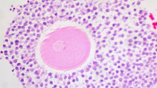
Picture: Ed Uthman via Flickr
A human egg is enormous. OK, it’s tiny when you compare it to a chicken egg, but at a shade over a tenth of a millimetre in diameter, it’s easily one of the largest cells in the human body. Its gigantism is particularly noticeable when you compare it to a human sperm. Or to the 25 sperm cells you can line up across its centre.
But the length differences between egg and sperm pale in comparison to their differences in volume. If a sperm head was the length of an average person, it would be trying to drill into an egg a bit bigger than the largest tank at the Georgia Aquarium. The one they keep the whale sharks in.
That’s a hell of a size difference, especially when you consider that both cells contain essentially the same mass of chromosomes. And that’s the important thing, right? Fusing the egg and sperm nuclei and pairing up mum and dad’s 23 chromosome contribution to make a new human genome? If that’s the case, what’s filling up the rest of that ten-thousand fold space difference inside the egg?
Surprisingly, most of it isn’t food. It’s true that chicken eggs and their ilk contain a huge food store, but we don’t develop on our own. Human embryos get to mooch off mum as soon as they implant in a uterus, typically about a week after fertilisation. There’s enough food to take the embryo through those early days: amino acids to build proteins, fats to make new cell membranes, and sugars to burn for energy, but there’s not a lot of it.
Then what is taking up all that space inside a human egg? Here are some of the things we’ve identified so far.
Initial Operating Instructions
Long strands of RNA sit dormant inside the ovum, arranged in orderly lattices, waiting to be activated by fertilisation. Every major type of RNA is there; all of it copied from the parts of the maternal genome important for early development. Strands of ribosomal RNA (rRNA) will become part of the cell’s protein-making machinery, forming ribosomes to read mRNAs and build proteins from amino acids. Transfer RNAs (tRNAs), will fold into shapes that can capture amino acids to carry them to the ribosomes. Messenger RNAs (mRNA) hold codes for the enzymes that activate proteins as they leave their ribosomal construction sites and other proteins important for cell survival and cell division.
Without the RNAs tucked into the oocyte by mum, the egg can’t develop after it’s fertilised. The new zygote will have a full compliment of chromosomes and two copies of every relevant gene, but its DNA is super-condensed: wound up and packed so tightly that DNA-reading proteins can’t attach to the DNA strands to access the code. In humans, the chromosomes don’t uncoil enough to be read until the embryo has between 4-to-8 cells. So all of the cell activity after fertilisation and the embryo’s critical first two cell divisions essentially need to run on autopilot.
The RNA instructions stored in the oocyte reactivate the dormant egg after fertilisation, help its nucleus fuse with the sperm’s, and guide it through its first two cell divisions. Most of it disappears once the embryo starts reading its own DNA.
Directions for Transformation
There are other kinds of instructions, too. Some mum-made mRNAs have been stashed in different parts of the egg, arranged in a way that will put them in different cells once the embryo starts to divide in earnest. These mRNAs code for proteins called morphogens.
Morphogens are like orchestra conductors: they tell the cells inside a developing embryo what kind of cell they need to become. They work by interacting with the DNA inside a cell, directly or indirectly, turning specific genes on or off, making the cell change position inside the embryo, or telling specific cells to divide or die. But they don’t behave the same way with every cell they meet.
Morphogen behaviour is concentration-dependent. That means they tell cells slightly different things depending on how much of the molecule a cell is exposed to. Morphogens are most concentrated in the cell that makes them, and they get less concentrated further away from that source. That behaviour means that different parts of the embryo get different instructions depending on their position.
As a result, morphogens are critical for telling embryonic cells, that are initially all the same, how they’re going to be different. The embryo will eventually make its own, but the morphogens provided by mum help define the most basic shape of the embryo: head vs. tail, front vs. back.
Tiny Little Powerhouses
Cell divisions take energy. Some of the machinery for getting that energy out of the food larder is stored in the mRNA codes, but most of it depends on mitochondria: a part of the cell with its own unique genes.
You may have already heard some of the story about mitochondria — bacterial origin, ability to use oxygen to make lots of chemical energy, symbiotic relationship with our cells — but you may not have thought through the implications. Mitochondria have their own genes, so they have to reproduce themselves as our cells divide. Cells without mitochondria don’t work correctly. And sperm mitochondria, for reasons we don’t understand, are usually destroyed after fertilisation. So when the egg develops, it’s mum that gives us a set of mitochondria to help fuel all our days and nights.
The “Interstates” of the Cell
The egg isn’t like a giant water balloon where everything floats around randomly. It’s organised, and its internal structures are interconnected by a network of membranes and protein struts. These components act like highways, moving proteins and RNAs from one regional hub to the next, sometimes modifying them on the way to make them more useful.
And a Protective Coating on Top
Over it all, a shell of gelatinous material sits just under the outermost membrane that surrounds the egg. The layer is filled with structures called cortical granules: packages of enzymes, adhesive molecules, and other proteins sealed in membranous wrappers that keep them separate from the rest of the cell. They stay sealed up until the first sperm pokes its way into the egg.
Moments after fertilisation, the cortical granules fuse with the egg’s outer membrane and spew their contents into the environment. The chemicals from the granules react with the gel surrounding the egg and make it inhospitable to other sperm. The change keeps additional sperm (and their now extraneous chromosomes) out of the egg, preventing a condition called polyspermy. Human embryos with multiple sets of chromosomes don’t develop correctly (and usually abort spontaneously), so in this case, more is most definitely not better.
Whatever your relationship with your mum, there is one thing to thank her for. [Alberts et al. 2002 [Bukovsky et al. 2005 [Simon and Pellicer (eds) 2009 [Gosden and Lee 2010 [Liu 2011 [Khalili et al. 2012 [Mueller et al. 2015]]