
Human flesh is opaque. As any good dictionary will tell you, that means it has the quality of “not transmitting light; being impenetrable to sight.” Well, forget that: scientists now can use light to see inside objects that were traditionally off-limits to the human eye — including our bodies.
Of course it’s already possible to use X-rays, MRI, ultrasound and the like to peer inside human beings, but results are never as crisp and clear as those acquired using visible light imaging. Not only that: optical wavelengths also interact with organic molecules — the one we’re made of — so visible light could also contain vital information about the tissue it travels through. It might reveals abnormalities in cells, say, or use information about bodily functions — something that other imaging techniques, such as MRI, resort to complex chemical tracers to achieve. And, perhaps most importantly, it’s also non-ionising, which is to say that, unlike X-rays and MRI, it doesn’t increase cancer risk at the intensities used for imaging.
Incoherent imaging
But sending light through something opaque is a challenge. Send a pulse of light into the human body and most of it’s reflected from the surface — that is, after all, what allows us to see our skin. But as anyone who’s shouted through a double-glazed window will know, just because most of the energy is reflected — in that case, as an echo — a little of it invariably propagates through the surface. It’s just hard to make out what. Sadly, when shining light into tissue, the attenuated signal that does make it through still has other barriers to contend with, as cells absorb and scatter it at every step. It’s not impossible to capture information from the scattered light — as we’ll find out — but it is difficult. Easier, instead, to make use of what light is reflected back directly.
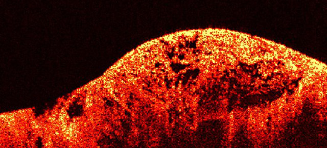
In the early 1990s, a technique known Optical Coherence Tomography used a phenomenon called interferometry to generate images. Light from a single sources was broken in to two arms: one kept as a references, the other shone at a sample. Combining the reflection from the light shone at the sample with that in the reference arm created an interferences pattern, but only for a single time of flight, where the light had traveled the same optical distance. By scanning a mirror up and down the reference arm, it was possible to map the interference at all depths for a single point. Move the light beam on the sample along to the side, then again, and again, and you create a 2D images, such at the one above (that’s a sarcoma on human skin). Move the light in another axis, too, and you create a 3D images, such as the one below (that’s a finger print; check out the grooves).
Only, the technique’s limited to imaging tissue at depths of less than 1 millimetre below the surface. At greater depths, too much light is lost, turning the image into junk. Still, those distances are certainly enough to image the top layers of the skin, and technique’s also found sensible applications in opthalmic circles to image the retina — because, and this may not surprise you, shining light through the eye is quite easy, actually.
Sound and vision
Bump up the intensity and change the light source, though, and some interesting things happen. In the early 2000s, researchers realised that they could image structures that seem opaque by pumping laser light pulses into biological tissues. Unlike Optical Coherence Tomography, the light here isn’t reflected or even scattered in a fathomable fashion — rather, it is absorbed by the tissue. In turn, that section very briefly warms up, expands, cools and contracts, in the process generating a change in pressure which manifests itself as a high frequency sound pulse. In other words, the injected laser light turns the body itself into a kind of ultrasound source — and by measuring the sound that’s emitted, it’s possible to build an image of the tissue’s structure.
Fortunately the frequency of the sound emitted is relatively high — in the order of ten of megahertz — which provides a resolution of the order of tens of microns. Not fine-grained enough to see anything at the sub-cellular level, but certainly enough to understand what’s happening within the body — to spot tumours, say. Perhaps most compelling is the fact that it can be used to see at depths that genuinely start to make it a useful and viable imaging technique in a medical setting — of at least 3mm and perhaps up to centimetres. Because of the way blood absorbs light — much more readily than most of the tissue in your body is how — photoacosutic imaging has found some success in imaging blood vessels. The image below, for instance, shows a melanoma in a mouse and the vasculature which surrounds it.
Let there be light
But we digress. Photacoustic imaging isn’t really seeing into the body with light — it’s merely using light to kick-start a process. In recent years, there’s been as attempt to use the light that’s scattered, not reflected or absorbed, to image what lies within opaque samples. “Our technique relies on the fact that, even if it is completely opaque, the scrambled field generated by an luminous object, which seems completely and hopelessly random, does contains some information about this object,” explains Sylvain Gigan, a physicist at the Kastler Brossel Laboratory in Paris. “Under some conditions, we showed that we were able to retrieve it, using clever algorithms and thanks to the deep understanding of the scattering process.”
The technique Gigan alludes to, explains a recent article in Nature, borrows from theories recently developed by astronomers. They have been working out how to remove distortion in images of stars, that’s created as light is scattered by the atmosphere on its journey to the telescope lens. There, an understanding that a star should appear as a single bright spot is used to work out how the atmosphere has scattered light, and an algorithm used to correct for the effects. In 2008, a team showed that a spatial light modulator — a device that could steer a laser beam by delaying part of it relative to another — could pump light into an opaque object, and some of the scattered light captured by a detector on the other side. Using knowledge of how it was delayed when it was transmitted and that it was a bright, single spot originally, it was possible to use the detected light to build up an image of subtle variations in the opaque barrier that would give rise to the scattering — and hence image it.
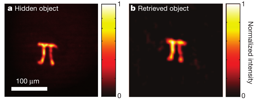
More experiments have followed, with the primary aim being to shift the detector so that it sits alongside the transmitter — making it potentially more useful for peering into the human body. That’s made possible by the fact that photons bouncing around inside a sample provide their own, weak illumination of their surroundings, which can in turn be reflected by a structure and received by the detector. That’s certainly been shown to work for florescent objects hidden in thin opaque samples — in this case, a π symbol — by scanning a laser over the surface of a sample and then using algorithms to reconstruct an image of what lies beneath. Gigan has even taken that work further, being able to achieve similar results but now in a single shot.
The dark before dawn
It’s still, arguably, early days for these techniques. “There is still a lot of room for new physics and important technical progresses to make it a reality,” explains Gigan. Indeed, other research groups are using similar thinking but different techniques to achieve similar ends. At Caltech, Professor Changhuei Yang is using ultrasound to induce a traceable frequency shift in laser light, that makes it possible to build up a map of scattered light by shifting the focus of the sound. Lihong Wang, a biomedical engineer at Washington University in St. Louis, has even used similar techniques to accurately image a piece of stained gelatin beneath a mouse ear.
Progress is certainly been made. But even if seeing inside the body with light doesn’t quite work out, it may even not matter. “I think going forward in the near future, imaging is not necessarily the primary goal,” explains Yang . “The ability to freely focus light deep in tissues enables one to start ablating tissues without harming superficial tissue layers. It also enables deep tissue biochemical analysis for disease diagnosis.” Regardless of how the research pans out, then, there’s a bright light at the end of this particular tunnel.
Picture: Soreen D/Flickr