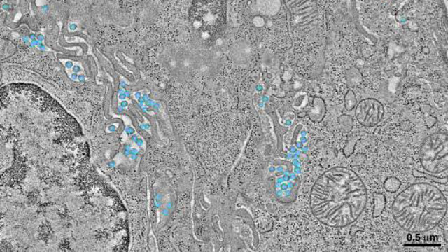
This monochrome image of living tissue has some extremely unwelcome visitors lurking within it. Taken from some of the first ever 3D images of HIV at work, those little blue circles show the virus infecting the surrounding cells.
The electron microscope images show HIV-1 virions in bright blue, embedded in the gut tissue of a lab mouse. Those mice were specially created by biologists from California and Massachusetts for the purposes of the research. That process involved transplanting human bone marrow, liver and thymus cells into the mice — in turn providing them with some of the immune cells that HIV attacks in people.
Then, the researchers infected the mice with HIV, left them for 10-to-20 weeks, and dissected them in order to inspect their guts and assess how HIV spreads through the body. Analysis by electron microscopy at different angles then allowed them to create 3D images of the virus at work.
The results, published in PLOS Pathogens, show that HIV seems to cluster in pools deep within the intestine, multiplying in peace, well away from immune cells. The images also provide more evidence to explain how infected cells spread HIV to neighbouring cells they touch — a process called virological synapse — which is known to happen but currently poorly understood. It’s yet another piece of research which will help in the battle against HIV. [California Institute of Technology via Popular Science]